Posterior-Anterior (PA) View Chest Radiograph.
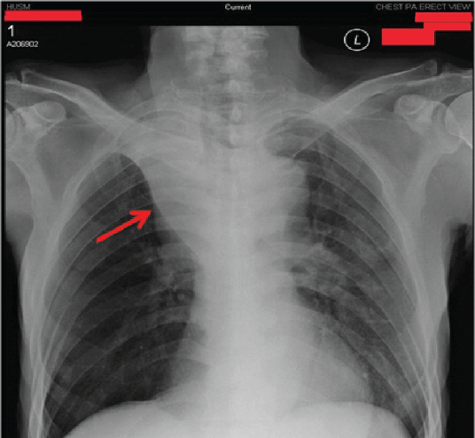
Legend: Figure 1 shows a right upper lobe collapse (red arrow) with loss of pulmonary vascular markings. The trachea also deviated towards the right side.
Zhi Xiong Chong1, Nazmi M. Noori2
doi: http://dx.doi.org/10.5195/ijms.2015.117
Volume 3, Number 1: 55-58
Received 26 09 2014: Accepted 19 01 2015
ABSTRACT
Background:Small cell lung cancer is an aggressive subtype of lung cancer whereby about one-third of cases are complicated with brain metastases. However, cerebellar metastases are uncommon and contribute to less than 10% of brain metastases.
Case:We report a 76-year-old Malay male, an active smoker who presented with dyspnea and occasional cough with hemoptysis for one week. He also pre sented with headache and constitutional symptoms of malignancy. Clinical examination suggested the presence of right upper chest patholo gy and positive left cerebellar signs. His condition deteriorated two days later and he passed away after failed attempts at resuscitation. Chest radiograph showed right upper lobe collapse, and brain magnetic resonance imaging showed metastatic lesion in the left cerebellum extending to the right cerebellum. Post-mortem findings revealed small cell lung cancer with cerebellar metastases.
Conclusion:Small cell lung cancer patients with brain metastases deteriorate very rapidly, and the management is mainly supportive. Primary prevention through education is the best way to reduce the incidence of lung cancer. In addition, secondary prevention and screening should be undertaken at earlier stages of the disease, as some studies have shown that combined chemotherapy and radiotherapy improve prog nosis of malignancies detected at early stage.
Keywords: Lung Neoplasms; Small Cell Lung Carcinoma; Neoplasms Metastasis; Cerebellar Neoplasms (Source: MeSH, NLM).
Lung cancer can be divided into two types, small cell (SCLC) and non-small cell carcinomas (NSCLC).1 SCLC makes up about 20% of all lung cancer cases.1,2 Compared to NSCLC, SCLC has a more aggressive disease course and almost always metastasizes to extra-thoracic organs.2,3 70% of patients with SCLC present with extra-thoracic metastases at the time of diagnosis.3 Brain metastases are seen in 30.5% of patients with SCLC.1,4 Cerebellar metastases are uncommon and contribute to less than 10% of brain metastases.1,2,5
Essentially, the prognosis of the SCLC patient with brain metastases is very poor with a median survival of one year.3,5 One isolated study has reported survival of more than three years following intensive combination of high dose chemotherapy and radiotherapy.3 In advanced cases, palliative care is as important as chemotherapy to improve the quality of life of the patient.5
We report an advanced case of SCLC with brain metastases, the management of which was mainly supportive. We would like to highlight the importance of primary and secondary prevention in the management of SCLC, as advanced disease carries a poor prognosis.1,5
Written informed consent was obtained from both the patient and his wife to discuss his case in the form of a published case report.
This is a case of a 76-year-old Malay male farmer, an active smoker who started to smoke 50 years ago with a 40 pack-year history. He developed a non-productive cough six months prior to hospital admission. Two months later, he developed a headache, nausea, vomiting and loss of appetite. His original weight was 80 kilogram, of which he lost 15 kilogram (or 19% of his body weight). Initially, his daily activity remained normal. However, one month prior to admission, he developed difficulty in maintaining his balance while walking and became dependent on activities of daily living.
One week prior to admission, he started experiencing dyspnea and an exacerbation of his cough. On the day of admission, he developed hemoptysis and severe dyspnea, not relieved by rest. He was brought by family members to the hospital. On arrival, he was alert and was given oxygen support via nasal prong at 3L/minute. He had no symptoms of heart failure, chest pain, fever, history of tuberculosis contact, night sweating, history of prolong immobilization, calf pain or hematemesis.
The patient has underlying hypertension and hyperlipidemia diagnosed two years earlier and is on regular follow-up. His last follow-up was six months ago and he did not complain of any problems to his primary care physician. Cardiovascular and respiratory examinations were normal during that visit. He is compliant with his medication (perindopril 2mg and atorvastatin 20mg daily). He has no family history of malignancy.
On physical examination, he was conscious, in pain and in respiratory distress. His blood pressure was 130/88mmHg. Respiratory examination revealed right upper lung pathology evidenced by reduced chest expansion and vesicular breath sounds. Bilateral supraclavicular lymph nodes were palpable. Cerebellar signs such as dysdiadochokinesia, positive finger nose test and heel shin test were present and predominantly affected the left side of his body. Examinations of other systems were unremarkable.
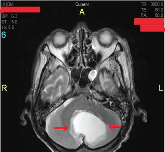
A series of investigations were completed. Arterial blood gas (ABG) demonstrated type I respiratory failure with PaO2 of 53mmHg. A full blood count and liver function tests were normal. Blood urea and serum electrolyte showed a slight elevation of serum urea and creatinine. The chest radiograph revealed a right upper lobe collapse with loss of pulmonary vascular markings (Figure 1). T2-weighted MRI displayed a single ill-defined hyperintense lesion measuring 6 × 5 cm in the left cerebellum extending to the right cerebellum (Figure 2). His electrocardiography (ECG) showed sinus rhythm and was normal. Tuberculosis work-ups, including a Mantoux test and sputum acid-fast bacilli were negative.
Figure 1.Posterior-Anterior (PA) View Chest Radiograph.
T2-weighted MRI of the Patient.
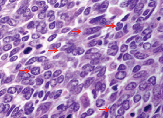
Bronchoscopy was performed and demonstrated the presence of a mass in the right main bronchus measuring 5 × 3 cm. Biopsy confirmed small cell lung cancer (SCLC) as the histological diagnosis (Figure 3). Computerized Tomography (CT) scan of the thorax was not completed, as the service was not available at that time. Based on the examination findings, bronchoscopy and MRI, he was diagnosed with stage IV (T2N3M1) SCLC with cerebellar metastases.
Figure 3.Higher Magnification of the Small Cell Lung Cancer Showing Numerous Mitotic Figures (red arrows).
The possible differential diagnoses included pulmonary and cerebellar tuberculosis, pulmonary embolism, stroke and myocardial infarction. Tuberculosis is common in Malaysia, however he denied any night sweats, tuberculosis contact and tuberculosis work-ups were negative. Pulmonary embolism was possible, but he did not complain of pleuritic chest pain or have a history of immobilization. Stroke and myocardial infarction were possible based on his risk factors, such as male gender, old age, smoking, hypertension and hyperlipidemia, but the progressive onset of the disease made this unlikely. In addition, the ECG ruled out ischemic changes. Therefore, the most likely provisional diagnosis at this point was right lung cancer with cerebellar metastases.
He was admitted to the respiratory care unit and given oxygen support via oxygen mask at 9L/minute. He was also given intravenous morphine 10mg daily dose for pain relief and intravenous pantoprazole 20mg twice daily dose to prevent gastric ulcers.
He was scheduled for palliative combined chemotherapy and radiotherapy. He was counseled on the disease burden and the long term complications of the treatment. He understood and agreed to the treatment plan. The chemotherapy regimens included intravenous cisplatin 60–80mg/m2 on day 1 for every 28 days and intravenous etoposide 80–120mg/m2 on days 1 to 3 for every 28 days. Palliative low dose radiotherapy of about 20–30Gy was scheduled to be given in 5 to 10 fractions. Surgery was not indicated as the disease had already progressed to an advanced stage.
However, these therapies were not given because his condition rapidly deteriorated two days later. He collapsed suddenly, was intubated and given mechanical ventilation. Due to the sudden nature of his collapse, consent from the patient himself was not obtained for resuscitation. The decision to intubate was made after a discussion with family members. He was given intravenous adrenaline 1mg every five minutes. He passed away after failed cardio-pulmonary resuscitation (CPR) for 30 minutes. He was sent for post-mortem examination and autopsy confirmed that he had small cell lung carcinoma arising from right main bronchus with cerebellar metastases.
The rapid progression of SCLC that has metastasized to the brain has been well documented in the literature.1,5 Case reports report that patients may survive for three to five, after being treated with a combined chemotherapy and radiotherapy regimen.2,3
A systemic review and meta-analysis concluded that first line treatment for advanced SCLC should include four to six cycles of etoposide in addition to either cisplatin or carboplatin.6 Oral or intravenous topotecan is recommended for patients that are resistant to first line treatments.6 However, the routine use of thoracic irradiation in patients with metastatic SCLC is not recommended.6 The optimal surveillance of metastatic disease involves 3-monthly CT scans to monitor disease progression and response to treatment.6
A randomized control study showed that thoracic radiotherapy in addition to prophylactic cranial irradiation improved survival rates in the thoracic radiotherapy group than in the control group.7 However, Hirofumi Sakurai and et al., described two cases of SCLC with no metastatic lesions, where patients were started on early chemo radiotherapy and metastates were detected one and a half years later.8 This draws a question as to whether early treatment will benefit the patient. Generally, most of literature still supports early detection and treatment to reduce morbidity and mortality.1,2,5–7
The majority of published case studies only discuss the benefit of early detection and treatment.1,2,7 However, there is still a need for case reports which discuss a standardized management of advanced SCLC with brain metastases. Some case reports highlight the role of gamma knife surgery and intracranial irradiation in advanced disease to prolong the survival rate.9,10 However, most patients do not easily accept brain surgery and irradiation because of concerns of intra-operative complications and post-operative intellectual reduction.7 As for chemotherapy regimens, combination of cisplatin and etoposide are usually used although it still has limited proven efficacy in preventing metastatic disease.7,8 All these factors contribute to the limited success rate in the treatment of advanced SCLC.8
In conclusion, the rapid progression of advanced SCLC has poses many difficulties to clinicians treating this cohort of patients. Although there is no definitive treatment for advanced SCLC, we would like to stress the benefits of prevention and early detection.11,14 Attention should be drawn towards smoking as one of the most important contributors in the pathogenesis of lung cancer.12,13 Primary prevention by education on smoking cessation should be implemented and enhanced in the healthcare system.13–15 Images of lung cancer and obstetric complications caused by smoking have been printed on cigarette packing of many countries to discourage smoking.14 A study has shown that public education on tobacco smoking is still very low and public education campaigns are needed urgently to raise the public awareness on the harmful effects of tobacco products.16 Secondary prevention, which includes lung cancer screening among active and past smokers, should be encouraged for early disease detection.13,17 Tertiary prevention has limited role in treating advanced lung cancer.2,5 Therefore, prevention is always better than cure!
Key Points:The author has no funding, financial relationships or conflicts of interest to disclose.
Conception and design the work/idea: ZXC. Collect data/obtaining results: ZXC. Analysis and interpretation of data: ZXC. Write the manuscript: ZXC. Critical revision of the manuscript: NMN. Approval of the final version: NMN. Contribution of patients or study material: ZXC. Statistical advice: NMN. Administrative or technical advice: NMN.
We would like to express deepest gratitude to the patient, LMDZ, and his wife, MFM, for their willingness to participate in this case study.
1.Hoffman PC, Mauer AM, Vokes EE. Lung Cancer. Lancet. 2000 Feb 5;355(9202):479–85.
2.Imai R, Hayakawa K, Sakurai H, Nakayama Y, Mitsuhashi N, Niibe H. Small cell lung cancer with a brain metastasis controlled for 5 years: a case re port. Jpn J Clin Oncol. 2001 Mar;31(3):116–8.
3.Jesien-Lewandowicz E, Spych M, Fijuth J, Kordek R. Solitary brain metastasis of an occult and stable small-cell lung cancer in a schizophrenic patient: a 3-year control. Lung Cancer. 2010 Aug;69(2):245–8.
4.Newman SJ, Hanssen HH. Frequency, diagnosis and treatment of brain metastases in 247 consecutive patients with bronchogenic carcinoma. Cancer. 1974 Feb;33(2):492–6.
5.Stupp R, Monnerat C, Turrisi AT 3rd, Perry MC, Leyvraz S. Small cell lung cancer: state of the art and future perspectives. Lung Cancer. 2004 Jul;45(1):105–17.
6.Früh M, De Ruysscher D, Popat S, Crinò L, Peters S, Felip E; ESMO Guide lines Working Group. Small-cell lung cancer (SCLC): ESMO Clinical Practice Guidelines for diagnosis, treatment and follow-up. Ann Oncol. 2013 Oct;24(Suppl 6):vi99–105.
7.Saito Y, Hayakawa K, Mitsuhashi N, Nakajima N, Kato S, Nakazato Y, et al. Late relapse of small cell lung cancer treated with radiation therapy alo ne—case report. Lung Cancer. 1994 Mar;10(5-6):319–24.
8.Sakurai H, Kurishima K, Homma S, Kagohashi K, Miyazaki K, Kawaguchi M, et al. Isolated solitary brain metastasis as a relapse of small cell lung cancer. Oncol Lett. 2013 Oct;6(4):1108–10.
9.Elaimy L, Mackay AR, Lamoreaux WT, Fairbanks RK, Demakas JJ, Cooke BS, et al. Clinical outcomes of stereotactic radiosurgery in the treatment of patients with metastatic brain tumors. World Neurosurg. 2011 May-Jun;75(5-6):673–83.
10.Pan HC, Sheehan J, Stroila M, Steiner M, Steiner L. Gamma knife surgery for brain metastases from lung cancer. J Neurosurg. 2005 Jan;102 Suppl:128–33.
11.Whittemore AS. Effect of cigarette smoking in epidemiological studies of lung cancer. Stat Med. 1988 Jan-Feb;7(1-2):223–38.
12.Miller YE. Pathogenesis of lung cancer: 100 year report. Am J Respir Cell Mol Biol. 2005 Sep;33(3):216–23.
13.Liu BQ, Peto R, Chen ZM, Boreham J, Wu YP, Li JY, et al. Emerging tobacco hazards in China: Retrospective proportional mortality study of one million deaths. BMJ. 1998 Nov 21;317(7170):1411–22.
14.Hann CL, Rudin CM. Management of small-cell lung cancer: incremental changes but hope for the future. Oncology (Williston Park). 2008 Nov 30;22(13):1486–92.
15.Burns DM. Primary prevention, smoking, and smoking cessation: implications for future trends in lung cancer prevention. Cancer. 2000 Dec 1;89(Suppl 11):2506–9.
16.Tan LM. Future of tobacco industry in Malaysia. The Star. 2014;11(200):2–3
17.van Iersel CA, de Koning HJ, Draisma G, Mali WP, Scholten ET, Nackaerts K, et al. Risk-based selection from the general population in a screening trial: selection criteria, recruitment and power for the Dutch-Belgian randomised lung cancer multi-slice CT screening trial (NELSON). Int J Cancer. 2007 Feb 15;120(4):868–74.
Zhi Xiong Chong, 1 Final Year Medical Student, School of Medical Sciences, Universiti Sains Malaysia (Malaysian Science University), Malaysia.
Nazmi M. Noori, 2 MBBS, PhD. Department of Medicine, School of Medical Sciences, Universiti Sains Malaysia (Malaysian Science University), Malaysia.
About the Author: Chong Zhi Xiong is currently a fifth year medical student of Universiti Sains Malaysia, Kubang Kerian, Malaysia of a five year program. He is also a winner in scientific poster competition in East Asian Medical Student Conference (EAMSC) in Japan in 2013.
Correspondence: Zhi Xiong Chong. Address: Universiti Sains Malaysia Health Campus, Jalan Raja Perempuan Zainab II, 16500 Kubang Kerian, Kelantan, Malaysia. Email: zhixiong17c@yahoo.com
Cite as: Chong ZX, Noori NM. Advanced Small Cell Lung Cancer with Cerebellar Metastases – A Case Report. Int J Med Students. 2014 Nov-2015 Mar:3(1):55-8.
Copyright © 2015 Zhi Xiong Chong, Nazmi M. Noori
International Journal of Medical Students, VOLUME 3, NUMBER 1, March 2015