Review
Ocular Auscultation: A Review
Daniel Fernando Gallego1, Ana Maria Rivas-Grajales2, Carlos Jose Gallego3
doi: http://dx.doi.org/10.5195/ijms.2015.125
Volume 3, Number 2: 102-106
Received 24 12 2014:
Accepted 31 03 2015
ABSTRACT
Ocular auscultation is a commonly neglected step of routine physical examination.
An adequate ocular auscultation can be helpful in discovering an ocular bruit, which
is an important diagnostic finding for a broad spectrum of pathologic conditions,
some of which are potentially fatal. In this article, we present a literature review
on the physical exam maneuver of ocular auscultation, as well as the pathophysiology
and differential diagnosis of ocular bruits. We also included a description of the
adequate auscultation technique and a discussion about the applicability of ocular
auscultation in clinical practice.
Keywords:
Auscultation;
Physical Examination;
Carotid Stenosis;
Carotid-Cavernous Sinus Fistula;
Neurological Examination.
Introduction
Ocular auscultation is the physical exam maneuver that consists of listening to the
vascular sounds of the head and neck by placing the stethoscope on the surface of
the eyelids and surrounding structures.1 The development of an ocular murmur is secondary to the turbulent flow inside the
vessels around the orbit, which can arise from localized pathologies (e.g. stenosis
of the carotid artery) or systemic conditions (e.g. anemia).2–5 Moreover, ocular bruits have been identified in patients suffering from life-threatening
conditions, such as subarachnoid hemorrhage, stroke, and carotid-cavernous fistulas.6–8 Ocular bruit has also been reported as the only auscultatory finding in cases of
symptomatic atherothrombotic vascular disease.9
Despite its clinical relevance, the auscultation of the orbit is often neglected in
the routine neurological examination, especially now that better diagnostic tools
are replacing clinical examination,1 including the use of Doppler ultrasound technology in evaluating orbital lesions.10 Anyhow, physical exam maneuvers and radiological tools are not mutually exclusive
and, in other scenarios, have been proven to have additive diagnostic efficacy. For
example, the use of cardiac auscultation complemented by echocardiography has shown
improved accuracy in murmur identification compared to echocardiography or physical
exam alone.11
The non-use of ocular auscultation in clinical practice could be due to the lack of
knowledge of the technique and the lack of awareness of the clinical implications
of an orbital bruit. In this article, we present the pathophysiology and differential
diagnosis of orbital bruits, as well as a brief description of the ocular auscultation
technique. We also included an Evidence Based Medicine section with a literature review
on ocular auscultation and the prevalence of ocular bruits in selected populations.
Pathophysiology of Ocular Bruits
An understanding of vascular hemodynamics is useful for the interpretation of vascular
sounds in any anatomical site. An arterial bruit indicates the presence of stenosis
at or proximal to the area of auscultation. As the stenosis increases, the potential
energy (pressure) proximal to the stenosis is transformed into kinetic energy (velocity)
within the stenosis, resulting in a turbulent flow and an audible sound. Cranial and
orbital bruits represent vibrations arising from vascular structures within the cranium,
neck and, occasionally, from cardiac lesions. The orbits serve as a “window” for sound
transmission and minimize dissipation through bony structures. There are four factors
that may alter the intensity and duration of arterial bruits: high inflow resulting
from a high cardiac output, diminished side-branch flow, poor or absence of collateral
vessels, and augmented outflow.2
Regarding ocular bruits, three underlying pathophysiological processes related with
the aforementioned factors should be suspected. First, the confluence of blood vessels
with high blood flow resulting in a high arteriovenous pressure difference in the
proximities of the ocular cavity; this is characteristic of vascular malformations
and carotid-cavernous fistulas, in which a considerable blood volume is diverted from
vessels with high hydrostatic pressure (arteries) to those with low hydrostatic pressure
(veins).12 Second, the occlusion in the internal carotid artery with subsequent ipsilateral
and contralateral arteriolar vasodilation; this is the case of stenotic lesions and
a flow deviation to contralateral vessels. Finally, an ocular bruit could be a sign
of increased cardiac output, as seen in anemia and hyperthyroidism.
Differential Diagnosis of Ocular Bruits
An ocular bruit can be associated with a wide range of pathologies;1 therefore, a thorough history and clinical examination is essential.13–14 Positive auscultatory findings should suggest these diagnoses only if the entire
clinical picture is supportive.2 The main conditions that have been associated with this finding are presented in
Table 1.
Table 1.
Differential Diagnosis of Ocular Bruit.
- Vascular conditions
- Carotid-cavernous fistula
- Arteriovenous malformations
- Cerebrovascular accidents
- Severe atherosclerosis
- Internal carotid artery stenosis
- Vasculitides
- Churg-Strauss disease
- Temporal artery vasculitis
|
2. Systemic conditions
- Anemia
- Thyrotoxicosis
- Paget's disease
|
3. Irradiation from distant structures
- Aortic aneurisms
- Aortic stenosis
- Hypertension (in infants)
|
Carotid-cavernous fistula is the main condition that should be suspected when an ocular
bruit is found in clinical examination. This finding is part of a diagnostic triad
consisting of proptosis, chemosis and ocular bruit and has been reported in 50% of
cases.15–16 In patients with vascular malformations, which can be silent despite their size,
an ocular bruit could be the only physical finding.4 The confluence of high-flow vessels is the underlying pathophysiology in this condition.
One other disease that is associated with ocular bruits is the presence of an ischemic
cerebrovascular accident or a transient ischemic attack due to stenosis in the internal
carotid artery.5–17 Two cohorts of patients with cerebrovascular disease reported a prevalence of ocular
bruits of 28% and 0.6%, respectively,18,19 while another cohort of symptomatic stroke patients reported a prevalence of 72%.9 In cerebral ischemia, the collateralization process determines the infarcted area.20 When the internal carotid artery is occluded, a retrograde flow deviation occurs
through the external carotid artery via the ophthalmic artery towards the intra-cerebral
system, producing the vascular murmur.21 Vasodilation of the episcleral arteries has been described as an additional useful
physical finding.22 A cautious palpation of the facial artery branches may reveal a hyperdynamic high-grade
lesion in the internal carotid artery.3
The vasculitides can also present with an ocular bruit as a consequence of vessel
incompliance due to systemic inflammation and possibly due to narrowing of the vessel
lumen. For example, patients with giant cell arteritis, which is characterized by
an inflammation in the lining of the temporal artery, can be associated with an ocular
bruit.23,24 This finding has also been reported in a patient with Churg-strauss syndrome.25
Conditions that increase the systemic blood flow (e.g. anemias) should be considered
in the differential diagnosis. The presence of an ocular bruit has been reported in
two case series with chronic kidney injury.26,27 Ocular bruits have also been described in Paget's disease, in which the increased
cardiac output results from an increased rate of angiogenesis.28 Finally, an ocular bruit can radiate from distant vascular structures, such as thoracic
and abdominal aneurysms, aortic stenosis, and hypertension in pediatric patients.14
Ocular Auscultation Technique
Auscultation should take place in a quiet room with both the patient and the examiner
relaxed and in a comfortable position. Historically, a large and narrow bell has been
used in ocular auscultation, like the one included in the Ford-Bowles stethoscope
(Figure 1). However, for practical reasons, the bell found in modern stethoscopes is considered
appropriate. Cranial bruits should be listened over the skull, and examination should
include the frontal, temporal and mastoid regions and the eyeball, with the latter
being more favorable for fainter sounds.2
Figure 1.
Ford-Bowles Stethoscope. Note the large and narrow bell that allows the identification
of murmurs while performing peripheral vascular auscultation. This modern version
is accompanied with a diaphragm to complement the auscultation of other systems for
a thorough physical examination.
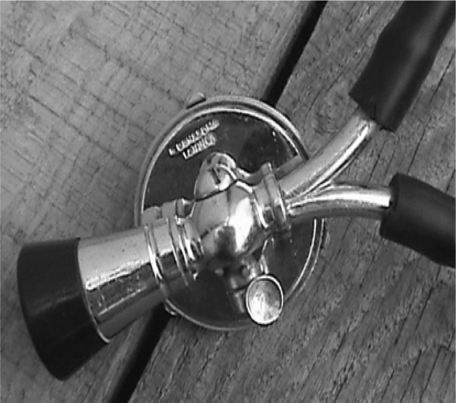
Legend: From Retonthenet Copyright©2008-2014 All rights reserved. Reprinted with permission
from David Skinner.
The auscultation of the orbit should be done by gently placing the bell of the stethoscope
over patient's closed eye (Figure 2). To minimize the sound produced by eyelid tremor, the patient should be asked to
stare at a fixed point while the examiner gently closes one of the eyes and firmly
places the stethoscope over the closed eye. If the patient is unable to keep his sight
fixed, the examiner can help by placing a finger as a reference point in front of
the patient's eyes. Finally, the patient should be asked to hold his breath. Orbital
bruits are usually faint and high-pitched, and the examiner should focus on the systolic
phase of the cardiovascular cycle. Placing a thumb over the carotid artery should
help in identifying the first heartbeat.13
Figure 2.
Adequate Technique for Ocular Auscultation. The bell of the stethoscope should be
placed over the patient's closed eye. The examiner's finger can be used to keep the
patient's sight fixed and avoid eyelid tremor (A). Auscultation should include the
zygomatic (B) and temporal regions (C).
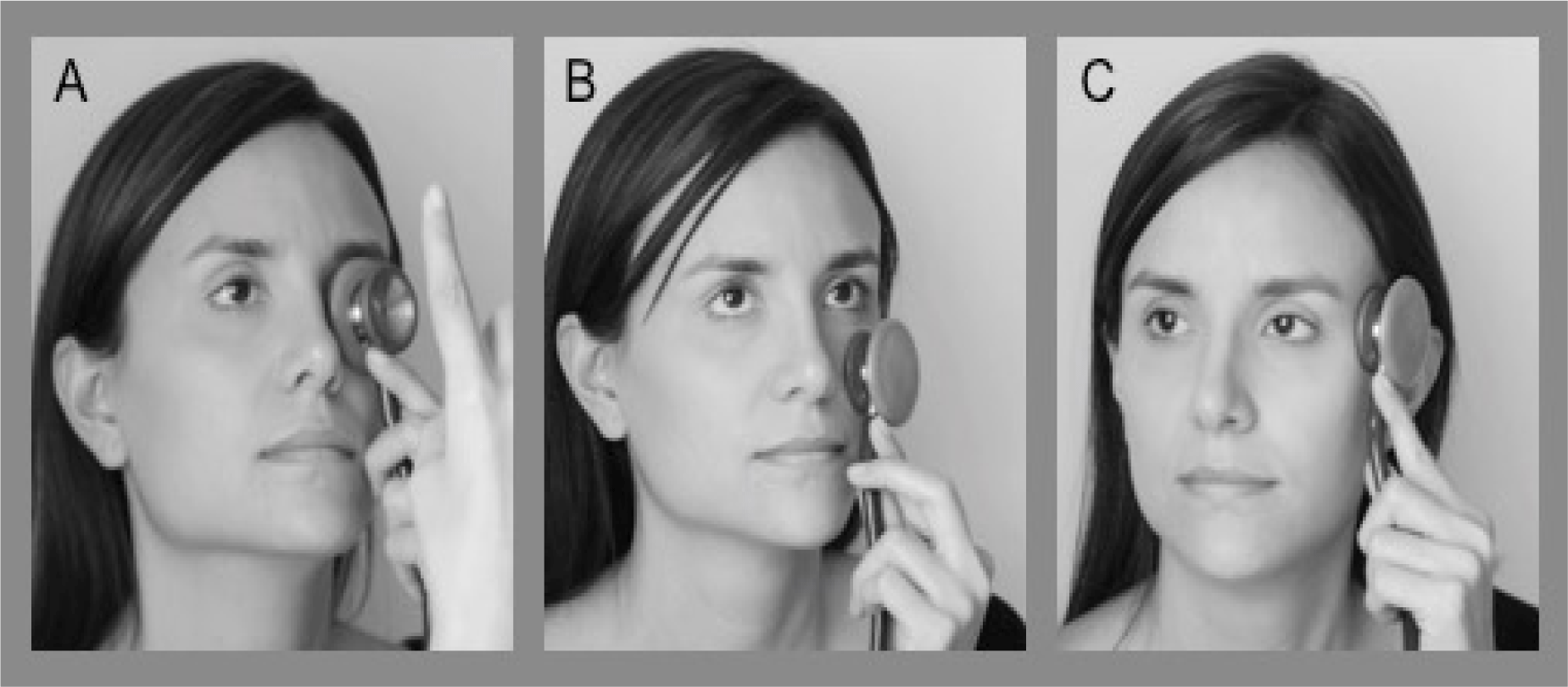
Auscultation of the frontal, zygomatic, temporal, and mastoid regions should be performed
using the diaphragm of the stethoscope and always be preceded by an adequate inspection
and palpation. Forced expiratory maneuvers, such as Valsalva maneuver, can be used
to intensify the bruit.2
Prevalence and Clinical Significance of Ocular Bruits: An Evidence Based Medicine
Section
A literature review was performed using PubMed/Medline, Embase and Scielo databases,
searching for the terms ‘ocular auscultation’, ‘ocular bruit’, and ‘ocular murmur’.
Additional relevant papers were retrieved from the articles' references. All reviewed
abstracts and articles were in English. The amount of literature found was scarce
as we expected; the majority of articles were case series and case reports. No recent
reviews on ocular auscultation were found. This limitation restricts drawing conclusions
on two important issues: the prevalence of ocular bruits in health and disease and
the clinical importance of ocular auscultation in medical practice.
In relation to prevalence, to date there are no specific studies published with this
purpose, and the current epidemiological data is derived from case series and prospective
cohort studies. The only reported data for ocular murmurs in healthy population is
as an innocent finding in 30%-60% of normal infants and children under six years of
age.2 The reported prevalence in pathological conditions varies widely across studies,
with cerebrovascular disease being the most studied. For example, Hirose et al. reported
seven ocular bruits in 250 patients,18 and Gautier et al., found only one ocular bruit in 150 patients with cerebrovascular
disease.19 In contrast, Hu et al., found 72 ocular bruits in 50 patients with symptoms of stroke
or transient ischemic attack.9 While a definitive deduction is not possible with all of the studies being prospective
cohort studies, we believe that the differences in the prevalence could be accounted
for by demographic variables, such as age, race and clinical factors. For instance,
Hirose et al. studied a sample of patients with cerebrovascular diseases of variable
severity. On the other hand, Hu et al., included only patients with symptomatic atherothrombotic
ischemic carotid disease, which suggests the presence of a more severe underlying
condition. Further studies with standardized inclusion criteria aimed at evaluating
the prevalence of ocular bruit in cerebrovascular disease and non-cardiovascular conditions
are needed.
Concerning clinical importance, there are contradictory views about the utility of
ocular auscultation as a routine practice. A report by the National Institute of Health
in 1975 concluded that ocular auscultation was of limited use due to its poor predictive
value in lesion localization and the severity estimation.29
However, in favor of ocular auscultation in specific clinical settings, Purcell reported
a patient who underwent enucleation after an ocular trauma. Ocular auscultation was
not performed during physical examination, and the patient suffered a near fatal bleeding
during the procedure due to a ruptured arteriovenous fistula. The author concluded
that this event could have been prevented by a complete ocular examination.6
Ocular bruits have been shown to be a crucial finding in guiding diagnostic evaluation.
Hu et al., conducted a prospective study in patients with symptoms of cerebrovascular
disease.9 They found that an ocular bruit was the only auscultatory finding in 28% of the patients.
In addition, ocular bruits were 57% more common than neck bruits in patients with
intracranial carotid artery occlusion. Smith et al., reported a patient with severe
carotid stenosis, which manifested clinically as limb-shaking transient ischemic attack.5 The finding of an ocular bruit in the neurovascular examination shifted the diagnostic
evaluation towards a vascular condition rather than a focal motor seizure.5 These two studies illustrate how the presence of an ocular bruit could inform the
clinician about the existence of vascular conditions and provide guidance towards
a successful diagnosis.
Finally, Atta et al., compared the clinical characteristics of a retrospective cohort
of patients with venous stasis orbitopathy. The study findings revealed that 30% of
patients with carotid-cavernous fistula had an ocular bruit, compared to 0% in the
non-vascular group. Ocular bruit was found to be the only significant physical finding
useful in differentiating carotid-cavernous fistula from other etiologies, mainly
compressive mass lesions.30
Based on the previous studies, we propose that ocular auscultation should be performed
in all patients with clinical suspicion of cerebrovascular disease and carotid cavernous
fistulas. Despite the limited evidence supporting the predictive value of ocular auscultation,
we believe that awareness of the clinical relevance of ocular bruits is an important
step towards encouraging research efforts in this field.
Conclusion
We presented the clinical relevant points of ocular auscultation, including ocular
auscultation technique, pathophysiology, and differential diagnosis of ocular bruits.
In spite of the improvement of diagnostic tools, clinical examination remains an important
aspect of clinical practice due to its low cost and wide accessibility. Ocular auscultation
is required in the detection of ocular bruits, a physical finding that can lead to
the diagnosis of a wide range of diseases, some of which are life-threatening.
Although the literature on this subject is scarce, we believe there is enough evidence
to suggest that it is important for physicians to acknowledge the role of ocular auscultation
in patients with suspicion of cardiovascular and neurological conditions, especially
atherothrombotic diseases and carotid-cavernous fistula. Further studies are needed
to document the prevalence of ocular bruits in the general population and selected
populations (e.g. patients with cerebrovascular disease).
Acknowledgments
The authors would like to thank Dr. Maria Eugenia Zuluaga and Juan Manuel Zuluaga
for providing Figure 2. We would also like to thank Mr. David Skinner for allowing the reproduction of Figure 1.
Conflict of Interest Statement & Funding
The author has no funding, financial relationships or conflicts of interest to disclose.
Author Contributions
Conception and design the work/idea, Collect data/obtaining results, Critical revision
of the manuscript, Approval of the final version, Contribution of patients or study
material: DG, AMRG, CJG
References
1. Wadia NH, Monckton G. Intracranial bruits in health and disease. Brain. 1957 Dec;80(4):492–509.
2. Kurtz KJ. Bruits and hums of the head and neck. In: Walker HK, Hall WD, Hurst JW, editors. Clinical methods: the history, physical, and laboratory examinations. 3rd ed. Boston: Butterworths; 1990.
3. Fisher CM. Cranial bruit associated with occlusion of the internal carotid artery. Neurology. 1957 May;7(5):299–306.
4. Jura E, Gołabek R, Kruszewska J, Kryst-Widźgowska T, Trzebicki J. [Giant arteriovenous angiomas of the brain with scant clinical manifestations]. Neurol Neurochir Pol. 1991 Jan-Feb;25(1):107–13.
5. Smith JH, Fugate JE, Claassen DO. Pearls & oy-sters: the orbital bruit: a poor man's angiogram. Neurology. 2009 Oct 20;73(16):e81–2.
6. Purcell JJ Jr. The effect of unsuspected carotid-cavernous fistula in enucleation. Am J Ophthalmol. 1979 Nov;88(5):946–8.
7. Brodsky LP. Auscultation of the human eye as a potential diagnostic aid. J Am Optom Assoc. 1980 Nov;51(11):1031–2.
8. Pessin MS, Panis W, Prager RJ, Millan VG, Scott RM. Auscultation of cervical and ocular bruits in extracranial carotid occlusive disease:
a clinical and angiographic study. Stroke. 1983 Mar-Apr;14(2):246–9.
9. Hu HH, Liao KK, Wong WJ, Teng MM, Lo YK, Chu FL, et al. Ocular bruits in ischemic cerebrovascular disease. Stroke. 1988 Oct;19(10):1229–33.
10. Nisbet RM, Barber JC, Steinkuller PG. Doppler ultrasonic flow detector: an adjunct in evaluation of orbital lesions. J Pediatr Ophthalmol Strabismus. 1980 Jul-Aug;17(4):268–71.
11. Stokke TM, Ruddox V, Sarvari SI, Otterstad JE, Aune E, Edvardsen T. Brief group training of medical students in focused cardiac ultrasound may improve
diagnostic accuracy of physical examination. J Am Soc Echocardiogr. 2014 Nov;27(11):1238–46.
12. Biousse V, Mendicino ME, Simon DJ, Newman NJ. The ophthalmology of intracranial vascular abnormalities. Am J Ophthalmol. 1998 Apr;125(4):527–44.
13. Poppen JL. Cranial bruit; its significance. Surg Clin North Am. 1955 Jun;Lahey Clinic No.:881–6.
14. Tiefert JW. Orbital auscultation. Am Fam Physician. 1978 Dec;18(6):117–20.
15. Ellis JA, Goldstein H, Connolly ESJr Meyers PM. Carotid-cavernous fistulas. Neurosurg Focus. 2012 May;32(5):E9.
16. Kato M, Ikegame Y, Toyoda I, Ogura S, Kitajima H, Yoshimura S, et al. Hemispheric laminar necrosis as a complication of traumatic carotid-cavernous sinus
fistula. Neurol Med Chir (Tokyo). 2009 Jan;49(1):26–9.
17. Ali S, Khan MA, Khealani B. Limb-shaking transient ischemic attacks: case report and review of literature. BMC Neurol. 2006;6:5.
18. Hirose Y, Yanagi T, Ito Y, Yasuda T. [Clinical significance of carotid and ocular bruits in cerebrovascular disease]. Rinsho Shinkeigaku. 1992 Oct;32(10):1081–6.
19. Gautier JC, Rosa A, Lhermitte F. [Carotid auscultation. Correlation in 200 patients with 332 angiograms]. Rev Neurol (Paris). 1975 Mar;131(3):175–84.
20. Ebihara A, Ashida T, Sugiyama T, Okuno S, Fujii J, Yonemitsu T. [An elderly hypertensive patient with ocular bruits and angiographically confirmed
stenosis of intracranical internal carotid arteries]. Nihon Ronen Igakkai Zasshi. 2002 Jan;39(1):94–6.
21. Lauritzen M, Alving J, Paulson OB. Orbital bruits and retinal artery pressure in internal carotid artery occlusion. Clin Neurol Neurosurg. 1981;83(1):7–10.
22. Countee RW, Gnanadev A, Chavis P. Dilated episcleral arteries––a significant physical finding in assessment of patients
with cerebrovascular insufficiency. Stroke. 1978 Jan-Feb;9(1):42–5.
23. Gilbert GJ. Physical diagnosis of temporal arteritis. Eye Ear Nose Throat Mon. 1971 Dec;50(12):476–8.
24. Gilbert GJ. Eyeball bruits in temporal arteritis. Dis Nerv Syst. 1970 Feb;31(2):130–2.
25. Jazayeri F, Pearson A. Orbital bruit in Churg-Strauss orbitopathy; a novel sign. Orbit. 2012 Apr;31(2):65–6.
26. Wales RT, Martin EA. Arterial bruits in anemia. Br Med J. 1963 Dec 7;2(5370):1444–7.
27. Parry DH, Worwood M, Jacobs A. Letter: Increased serum iron in acute leukaemia. Br Med J. 1975 Aug 9;3(5979):372.
28. Nugent JS, O'Brien KE Harris M, Mohan C. Paget's disease of bone in an Indian patient: genetic and environmental factors. J Clin Rheumatol. 2002 Aug;8(4):212–6.
29. National Institute of Health. A classification and outline of cerebrovascular diseases. II. Stroke. 1975 Sep-Oct;6(5):564–616.
30. Atta HR, Dick AD, Hamed LM, Byrne SF, Gendron EK, Hughes RL, et al. Venous stasis orbitopathy: a clinical and echographic study. Br J Ophthalmol. 1996 Feb;80(2):129–34.
Daniel Fernando Gallego, 1 Intern, Human Biology Division, Fred Hutchinson Cancer Research Center, USA.
Ana Maria Rivas-Grajales, 2 Graduate Student, Institute of Cognitive Neuroscience, University College of London,
UK.
Carlos Jose Gallego, 3 Acting instructor, Division of Medical Genetics, University of Washington, USA.
About the Author: Daniel Fernando Gallego is an intern at the Human Biology Division of the Fred Hutch
Cancer Research Center in Seattle, Washington, United States.
About the Author: Ana María Rivas-Grajales is a Graduate Student, Institute of Cognitive Neuroscience,
University College of London, UK.
Correspondence: Daniel Fernando Gallego, Address: 1100 Fairview Ave N, Seattle, WA 98109. Mail Stop
C1-015. Email: dgallego@fhcrc.org
Cite as: Gallego DF, Rivas-Grajales AM, Gallego CJ. Ocular Auscultation: A Review. Int J Med Students. 2015 Apr-Aug;3(2):102-6.
Copyright © 2015 Daniel Fernando Gallego, Ana Maria Rivas-Grajales, Carlos Jose Gallego
International Journal of Medical Students, VOLUME 3, NUMBER 2, August 2015