Case Report
Romantic Name for a Deadly Condition: Kissing Aneurysms of the Pericallosal Artery
– A Case Report
Przemyslaw M. Waszak1, Agnieszka Paturej1, Janusz Springer1, Katarzyna Baranowska1, Barbara Brzeska1, Katarzyna Aleksandrowicz1, Tomasz Szmuda2, Hanna Garnier1
doi: http://dx.doi.org/10.5195/ijms.2015.126
Volume 3, Number 2: 107-111
Received 09 12 2014:
Accepted 10 04 2015
ABSTRACT
Background:
Kissing aneurysms are two independent but adjacent aneurysms protruding from two contralateral
arterial locations. This report describes a successfully treated case of kissing aneurysms
at the Department of Neurosurgery, Medical University of Gdansk.
Case:
A 45-year-old asymptomatic woman was diagnosed with unruptured bilateral aneurysms
located in the pericallosal-callosomarginal division. Her medical history included
a previous intracranial aneurysm and arterial hypertension. The patient underwent
a successful treatment by surgical clipping and was discharged in good condition;
neither disability nor neurologic deficit was noticed upon discharge. Surgical wound
healing was complicated by an infection and resulted in a reoperation for the patient.
Conclusion:
The etiology of kissing aneurysms is still unknown and the best treatment method stills
remains unclear. Thus, every case has to be carefully and individually assessed by
an interdisciplinary team. As a result, patient transfer to an experienced neurosurgical
center could be beneficial.
Keywords:
Intracranial Aneurysm;
Anterior Cerebral Artery;
Microsurgery;
Angiography;
Kissing aneurysms.
Introduction
The prevalence of intracranial aneurysm is estimated at approximately 3.2%.1 The major characteristics of aneurysms include type (saccular, fusiform, dissecting,
mycotic, blood-blister-like, distal, etc.), size (micro, small, medium, large, giant
etc.) and location (branching sites of anterior, medial or posterior cerebral artery
etc.). Unruptured aneurysms are asymptomatic in most cases. When ruptured, they cause
subarachnoid hemorrhage (SAH), also known as hemorrhagic stroke. Main SAH symptoms
include sudden onset of severe headache, seizures, and neurologic deficits with rapid
deterioration leading to loss of consciousness. Basic diagnosis involves computed
tomography (CT) imaging or lumbar puncture. SAH represents a state of medical emergency;
even when treated early, SAH is associated with mortality up to 50% (including neurologic
deficits in many of the survivors).2 The main treatment options include open surgery with direct microsurgical clipping
of the aneurysmal neck or an endovascular procedure that occludes the aneurysmal lumen.
The risk of an aneurysm rupture depends on various risk factors such as the aneurysms'
characteristics (localization, type and size) and the patients' characteristics and
co-morbidities (hypertension, gender, cigarette smoking, alcohol intake, and prior
history of aneurysm).3,4
Kissing aneurysms (KAs) are unusual locational phenomena of multiple aneurysms. Although
the prevalence of multiple aneurysms can be up to 20% of all intracranial aneurysms,
the KAs – adjacent bilateral aneurysms arising from the same artery – are quite unique
with an incidence as low as 0.2%.5 Kissing aneurysms can be classified into two categories: type 1 represents aneurysmal
necks that are located on the same parent artery and type 2 exists where each aneurysmal
neck is located on different parent arteries.6 The most common location for KAs is the internal carotid artery (ICA), but other
sites such as the distal part of the anterior cerebral artery have also been reported.7 However, KAs associated with the posterior arterial supply of the brain are mainly
type 1. Kissing aneurysms are more common among women, especially middle-aged (40-59
years old).8 An interesting, but separate phenomenon is mirror-like (or twin) aneurysms that are
located bilaterally on analogs of arteries (eg. left and right middle cerebral artery,
MCA).9
Diagnosing KAs can be difficult in terms of determining the number or structure of
aneurysms based on radiologic examination. It has been suggested that more than half
of KAs are not been recognized preoperatively.6 Thus, the decision-making process can be particularly challenging. There is no recommended
screening for intracranial aneurysms, so in most of the cases they are revealed only
when they have ruptured. In a minority of the patients, the presence of an aneurysm
is known beforehand. In these special cases, it is highly important to make a prompt
diagnosis of unruptured aneurysms, evaluate the risk of bleeding, and consider its
eventual prophylactic treatment.
We present a case of unruptured KAs successfully treated surgically at the Neurosurgery
Department of the Medical University of Gdansk. Informed consent was obtained from
the patient for this study.
The Case
A 45-year-old asymptomatic woman was admitted to the Department of Neurosurgery for
the planned surgical clipping of unruptured bilateral aneurysms located in the division
of the anterior cerebral artery into the pericallosal and callosomarginal arteries
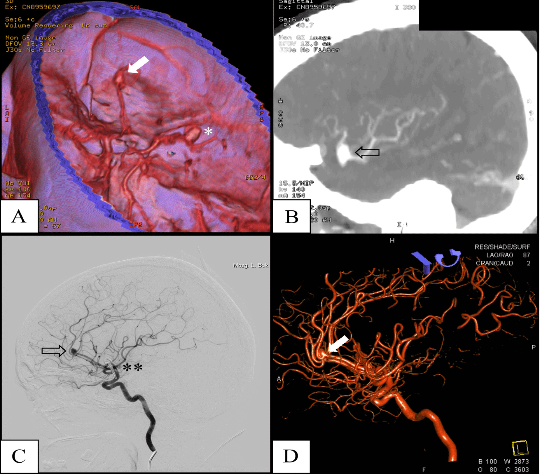
(A2/3 kissing aneurysms, Figure 1 A, B). Kissing aneurysms were radiographically diagnosed, after the patient's previous
aneurysm surgery.
Figure 1.
Radiologic Images of the Patient's Aneurysms.

Legend: A & B – Computed tomography angiogram (CTA) from 2012 showing the patient's aneurysms
at the division of the middle cerebral artery (MCA) into M1/M2 segments – 12 × 7 mm
(star) and at the bifurcation of the anterior cerebral artery to the pericallosal
and callosomarginal arteries (A2/3), so called kissing aneurysms – 4 × 4.5 mm (arrow);
A – a three-dimensional reconstruction, B – lateral CTA scan showing kissing aneurysms.
C & D – Digital subtraction angiography (DSA) from 2013 showing the same kissing aneurysms
(arrow) and a shade of the vascular clip from the previous MCA operation. C – DSA
showing the lateral presentation of aneurysms, D – a three-dimensional reconstruction.
The patient's medical history included hypertension, epilepsy, and 14 pack-years of
smoking. According to the patient's family, in 2012 she had an extreme case of alcohol
intoxication resulting in tonic seizures. Her surgical history included a previous
surgical clipping of an unruptured aneurysm in 2012 performed on the division of the
right middle cerebral artery (MCA) segments M1/M2. A computed tomography angiography
(CTA) scan was performed and revealed bilateral (kissing) aneurysms (4 × 4.5 mm in
size) occurring at the bifurcation of the anterior cerebral artery into the pericallosal
and calloso-marginal arteries (A2/3 division). This aneurysm was classified as type
1 as each aneurysmal neck was located on the same parent artery (Figure 1).
The patient's neurological status upon admission was normal and her general physical
examination was unremarkable. Cerebral angiography was performed and the diagnosis
of KAs was confirmed (Figure 1 C, D).
The patient underwent surgical clipping via a right frontal craniotomy approach under
general anesthesia. The skull was opened with a free bone flap. Opening of the interhemispheric
fissure was performed and the aneurysms were reached from the right and left side
(Figure 2). Clips on the necks of the aneurysms were applied without any complications. Vascular
patency was confirmed using ICG Pulsion (active ingredient: indocyanine green dye;
Pulsion Medical Systems SE); temporary clipping was not applied. No electrophysiological
monitoring was used during the surgical clipping. In addition, the surgery was prolonged
(5 hours).
Figure 2.
Surgical Clipping of Aneurysms – Intraoperative View.

Legend: A – The pia mater was carefully dissected and the pericallosal artery was exposed.
B – Both aneurysms at the bifurcation of the anterior cerebral artery into the pericallosal
and callosomarginal arteries are visible (arrows).
C – Subsequently, their necks have both been clipped.
D – Vascular patency was confirmed using ICG Pulsion (active ingredient: indocyanine
green dye).
The early postoperative course was uneventful. Magnetic resonance imaging (MRI) performed
on third post-operative day revealed no perfusion disturbances in the area of the
surgery.
The patient was discharged from the hospital on the 10th post-operative day in good
general condition; neither disability nor neurologic deficit was noticed (0 score
on the modified Rankin scale). No pre- or post-operative neuropsychological testing
was performed. The patient was referred for a follow-up appointment after 10-12 days
from the discharge date and a neurosurgical follow-up appointment after 8-10 weeks.
The surgical wound healing was complicated by an infection. As a result, the patient
received empirical clindamycin treatment. Given the wound infection, a reoperation
combined with cranioplasty was performed to evacuate the epidural pus and antibiotic
therapy was continued. This post-operative period was uneventful and the patient was
scheduled for a subsequent cranial allograft procedure.
Discussion
Kissing aneurysms derive their name from the specific spatial arrangement of two separate
but adjacent malformations. Besides certain congenital predispositions (e.g. Marfan
syndrome, Ehlers-Danlos syndome, autosomal dominant polycystic kidney disease etc.),
the causes and risk factors of KAs have not been fully defined.8 It can be assumed, however, that the risk factors are multifactorial and similar
to those of other types of multiple aneurysms. The described patient's gender, history
of smoking, hypertension, and an unclear episode of alcohol abuse should all be considered
as risk factors for aneurysm formation as well as contribute to the potential for
aneurysmal rupture in the future.8,10,11
Studies have suggested that 57% of KAs are not recognized preoperatively.6 Digital subtraction angiography (DSA) is the gold standard for detecting small and
large aneurysms. In non-emergency situations, DSA is essential to establish whether
acute treatment is needed or not, and if so, to select a proper treatment option.
Angiograms can, however, be misleading or even negative as described in a similar
report.7 Computed tomography angiography could also be a helpful tool to visualize such malformations
because it provides an option for the non-invasive imaging of KAS'. Importantly, it
should be emphasized that CTA can miss aneurysms smaller than 3mm or give false positive
results.7 Recently, more and more unruptured intracranial aneurysms are detected incidentally
using MRA but treatment decisions are rarely made based on MRA alone.
Management of unruptured aneurysm remains controversial.3,4 Generally, aneurysms smaller than 7mm are at a low rupture risk.12 However, lesions located in the anterior circulation are at an intermediate risk
of rupture.12 In this case, taking into consideration the patient's risk factors, the surgical
protection of KAs seems desirable. According to Rinkel et al., the relative risks
(RR) for aneurysm rupture and their corresponding risk factor in our patient included
hypertension (RR 2.0), heavy alcohol intake (RR 2.1), female gender (RR 2.1), smoking
(RR 3.4), and the presence of multiple aneurysms (RR 1.7).11 The multidisciplinary team decided upon the microsurgical clipping of the aneurysm
necks. According to guidelines, 45-year-old patients can benefit from surgical treatment.
Endovascular treatment seems to be more challenging in circumstances involving KAs
given the two vascular origins without a direct communication and the aneurysms at
the distal parts of intracranial vessels (such as A2/A3) being more difficult to treat
by coiling.
The preferred treatment method for a single unruptured aneurysm remains controversial;
thus, an unruptured KA makes the decision process even more complicated.3 Endovascular treatment has lower overall unfavorable outcomes but the patient's age
seems to be a crucial factor.13 Surgery-related morbidity and mortality are quite low among patients under 60 years
of age.13 However, endovascular procedures can lead to an increased risk of recurrence or retreatment
in this patient group.13,14 It is noteworthy that the surgical procedure provides the neurosurgeon with access
and space for more maneuvers within the surroundings of the bilateral lesion. Surgery
is difficult due to the dual arterial supply of KAs.5 The procedure should secure both aneurysms simultaneously; however, this maneuver
can lead to a higher risk of intraoperative bleeding.5
The patient had regular (every 6 months) follow-up care at the Neurosurgery Outpatient
Clinic. Computed tomography angiography examination was performed after 12 months,
showing complete aneurysm occlusion with no new pathologies. Although the re-treatment
ratio (short-term prognosis) after the successful surgical clipping is low, the patient
is at risk of aneurysm recurrence (long-term prognosis).14 So far (after 1.5 years of observation), the patient's follow-up remains uneventful.
Kissing aneurysms are a treatment challenge because of their unknown etiology, difficult
imaging diagnosis, and no officially stated treatment method. In our opinion, every
case has to be considered individually by an interdisciplinary medical team. In order
to assure positive outcomes, patients with this malformation should be referred to
an experienced neurosurgical center. Further studies are needed to allow for a better
understanding of this condition and the therapeutic options available to patients
with KAs.
Key Points:
- The preferred treatment method of a single unruptured aneurysm remains controversial,
thus an unruptured KAs makes the decision process even more complicated.
- Surgery is difficult due to KAs dual arterial supply. The procedure should involve
securing both aneurysms simultaneously, however this maneuver can lead to higher risk
of intraoperative bleeding.
- KAs still remain a challenge, because of their unknown etiology, difficult imaging
and no officially-stated treatment method.
Acknowledgments
None.
Conflict of Interest Statement & Funding
The author has no funding, financial relationships or conflicts of interest to disclose.
Author Contributions
Conception and design the work/idea: PMW, AP, KA. Collect data/obtaining results,
Analysis and interpretation of data: PMW, AP, KB, BB. Write the manuscript: PMW, AP,
JS, KB, BB. Critical revision of the manuscript: PMW, JS, KA. Approval of the final
version: PMW, JS. Administrative or technical advice: JS.
References
1. Vlak MH, Algra A, Brandenburg R, Rinkel GJ. Prevalence of unruptured intracranial aneurysms, with emphasis on sex, age, comorbidity,
country, and time period: A systematic review and meta-analysis. Lancet Neurol. 2011 Jul;10(7):626–36.
2. Koffijberg H, Buskens E, Granath F, Adami J, Ekbom a, Rinkel GJ, et al. Subarachnoid haemorrhage in Sweden 1987-2002: regional incidence and case fatality
rates. J Neurol Neurosurg Psychiatry. 2008 Mar;79(3):294–9.
3. Brown RD, Broderick JP. Unruptured intracranial aneurysms: epidemiology, natural history, management options,
and familial screening. Lancet Neurol. 2014 Apr;13(4):393–404.
4. Darsaut TE, Estrade L, Jamali S, Bojanowski MW, Chagnon M, Raymond J. Uncertainty and agreement in the management of unruptured intracranial aneurysms. J Neurosurg. 2014 Mar;120(3):618–23.
5. Baldawa SS, Menon G, Nair S. Kissing anterior communicating artery aneurysms: diagnostic dilemma and management
issues. J Postgrad Med. 2011 Jan-Mar;57(1):44–7.
6. Harada K, Orita T, Ueda Y. [Large kissing aneurysms of the middle cerebral artery: a case report––classification
of kissing aneurysms]. No Shinkei Geka. 2004 May;32(5):513–7. Jap
7. Alimohammadi M, Bidabadi MS, Amirjamshidi A. Bilateral “Kissing” Aneurysms of the Distal Pericallosal Arteries: Report of a Case
and Review of the Literature. Neurosurg Q. 2010;20(4):308–10.
8. Casimiro MV, McEvoy AW, Watkins LD, Kitchen ND. A comparison of risk factors in the etiology of mirror and nonmirror multiple intracranial
aneurysms. Surg Neurol. 2004 Jun;61(6):541–5.
9. Salunke P, Malik V, Yogesh N, Khandelwal NK, Mathuriya SN. Mirror-like aneurysms of proximal anterior cerebral artery: report of a case and review
of literature. Br J Neurosurg. 2010 Dec;24(6):686–7.
10. Vlak MHM, Rinkel GJ, Greebe P, Greving JP, Algra A. Lifetime risks for aneurysmal subarachnoid haemorrhage: multivariable risk stratification. J Neurol Neurosurg Psychiatry. 2013 Jun;84(6):619–23.
11. Rinkel GJ, Djibuti M, Algra A, van Gijn J. Prevalence and risk of rupture of intracranial aneurysms: a systematic review. Stroke. 1998 Jan;29(1):251–6.
12. Bijlenga P, Ebeling C, Jaegersberg M, Summers P, Rogers A, Waterworth A, et al. Risk of rupture of small anterior communicating artery aneurysms is similar to posterior
circulation aneurysms. Stroke. 2013 Nov;44(11):3018–26.
13. Lawson MF, Neal DW, Mocco J, Hoh BL. Rationale for treating unruptured intracranial aneurysms: Actuarial analysis of natural
history risk versus treatment risk for coiling or clipping based on 14,050 patients
in the nationwide inpatient sample database. World Neurosurg. 2013 Mar-Apr;79(3-4):472–8.
14. Corns R, Zebian B, Tait MJ, Walsh D, Hampton T, Deasy N, et al. Prevalence of recurrence and retreatment of ruptured intracranial aneurysms treated
with endovascular coil occlusion. Br J Neurosurg. 2013 Feb;27(1):30–3.
Przemyslaw M. Waszak, 1 Medical University of Gdansk, Students' Scientific Association, Neurosurgery Department,
Poland.
Agnieszka Paturej, 1 Medical University of Gdansk, Students' Scientific Association, Neurosurgery Department,
Poland.
Janusz Springer, 1 Medical University of Gdansk, Students' Scientific Association, Neurosurgery Department,
Poland.
Katarzyna Baranowska, 1 Medical University of Gdansk, Students' Scientific Association, Neurosurgery Department,
Poland.
Barbara Brzeska, 1 Medical University of Gdansk, Students' Scientific Association, Neurosurgery Department,
Poland.
Katarzyna Aleksandrowicz, 1 Medical University of Gdansk, Students' Scientific Association, Neurosurgery Department,
Poland.
Tomasz Szmuda, 2 Medical University of Gdansk, Neurosurgery Department, Poland.
Hanna Garnier, 1 Medical University of Gdansk, Students' Scientific Association, Neurosurgery Department,
Poland.
About the Author: Przemyslaw M. Waszak is currently a 6th (final year) year medical student of the
Medical University of Gdansk, Poland. He is also the founder and editor of the first
Polish scientific handbook for medical students entitled “Idea - Research - Publication”
Correspondence: Przemyslaw M. Waszak Address: Marii Skłodowskiej-Curie 3A, Gdańsk, Poland. Email:
p.waszak@gumed.edu.pl
Cite as: Waszak PM, Paturej A, Springer J, Baranowska K, Brzeska B, Aleksandrowicz K, Szmuda T, Garnier H. Romantic Name for a Deadly Condition: Kissing Aneurysms of the Pericallosal Artery – A Case Report. Int J Med Students. 2015 Apr-Aug;3(2):107-11.
Copyright © 2015 Przemyslaw M. Waszak, Agnieszka Paturej, Janusz Springer, Katarzyna
Baranowska, Barbara Brzeska, Katarzyna Aleksandrowicz, Tomasz Szmuda, Hanna Garnier
International Journal of Medical Students, VOLUME 3, NUMBER 2, August 2015

