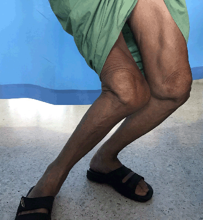
Knee Examination Bilaterally while Bearing Weight. Windswept Deformity towards the left of the patient.
Highlights:Yuki Julius Ng1, Kauseliya Velanthren2
doi: http://dx.doi.org/10.5195/ijms.2023.1513
Volume 11, Number 1: 71-75
Received 09 05 2022; Rev-request 05 06 2022; Rev-request 01 10 2022; Rev-recd 31 08 2022; Rev-recd 01 11 2022; Accepted 11 11 2022
ABSTRACT
Background:Twenty percent of the population globally is affected by musculoskeletal conditions. These conditions significantly impair mobility and dexterity. Pseudogout is similarly a debilitating disease that significantly increases morbidity and the disability adjusted life years of a person. We report a case of pseudogout in its advanced stage, causing total joint destruction of the knees and shoulders that manifested and presented as a windswept deformity.
The Case:Our patient is a 69-year-old man who complained of bilateral knee pain, shoulder pain during active flexion, and an obvious knee deformity. His familial history was not significant, and there was no history of injuries, infection, or congenital diseases. His knees were severely deformed, with extremely laxed collateral ligaments. Both of his shoulders had a limited range of movement with coarse crepitation on passive movement. X-ray of his knees showed a destroyed joint, reduced joint space, subchondral cysts, and chondrocalcinosis. X-ray of his shoulder joint showed a subluxated joint, subchondral cyst, and subchondral sclerosis. His joint aspirate was positive for rhomboid crystals in the birefringence test, consistent with pseudogout. Joint replacement surgery is the definitive management for this disease, but the patient and caretaker were not able to afford the implants.
Conclusion:We discussed the diagnosis of pseudogout in this patient and how the policies in place do not provide adequate coverage for these populations. This marginalizes those who need surgery and limits their access to affordable surgical care when needed.
Keywords: Chondrocalcinosis; Calcium Pyrophosphate Deposition Disease; Global Surgery (Source: MeSH-NLM).
One in five persons globally is affected by musculoskeletal conditions.1 These conditions significantly impair mobility and dexterity, causing early retirement and reducing the ability to participate in social activities. Pseudogout equally affects patients’ quality of life. Approximately 20% of patients with osteoarthritis requiring total knee replacement have calcium pyrophosphate deposition (CPPD) crystals in their joints.2 Although CPPD continues to be underdiagnosed, it is not difficult to confirm the diagnosis. Polarizing light microscopy with a red filter can accurately diagnose CPPD crystals. The hallmark of the crystal is its classical rhomboid shape and its relation to the light source. The crystal turns blue when the light axis is parallel and turns yellow when it is perpendicular under microscopy. This contrasts with monosodium urate crystals which turns blue perpendicularly and yellow parallel to the crystal axis in relation to the light source.3,4 The morphology of monosodium urate crystals appears as a needle-like rod-shaped crystals.4 We present a case of CPPD disease at its extreme stage of pathogenesis and discuss the diagnostic and social challenges faced.
A 69-year-old Chinese ethnic man with underlying hypertension presented with bilateral knee pain and windswept deformity, associated with right shoulder pain (Figure 1). He had no history of fever, numbness, leg weakness, trauma, or congenital anomaly. The knee pain started 3 years prior and his windswept deformity progressively worsened over the past 1 year. The bilateral knee pain was sharp in nature, did not radiate, and was exacerbated by walking and weightbearing. He scored his pain as 5/10. He was able to ambulate with a walking stick. His shoulder pain progressed over 8 years and was exacerbated with movement. He was primarily concerned with the prolonged nature of the pain. He was a construction worker before the deformity severely affected his joints.
Figure 1.Knee Examination Bilaterally while Bearing Weight. Windswept Deformity towards the left of the patient.
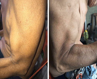
Upon examination, there was a large, boggy effusion of the shoulder joints bilaterally. The left shoulder had a reduced range of movement with gross crepitations during passive movement. Active abduction was up to 100° and frontal flexion was up to 90°. The right shoulder had a significantly reduced range of movements. Both active abduction and frontal flexion were up to 30°. The pain in both shoulders was localized with a pain scale of 5/10. Biceps bilaterally presented with Popeye deformity (Figure 2). Rotator cuff tests were abandoned due to the pain they caused.
Figure 2.Left and Right Popeye Deformity.
There was an obvious windswept deformity of the knee towards the left while bearing weight. Knee flexion and extension had a full range of motion on active and passive movement. Gross crepitus was felt with passive movement bilaterally. The left knee was able to passively angle medially up to 50° and the right knee was able to passively angle laterally up to 70° according to his deformity as shown (Figure 1). The knees were bilaterally tender upon performing the stress test with a pain scale of 5/10. McMurray’s test was inconclusive bilaterally due to the severely deformed anatomical structures.
His full blood count, renal profile, electrolytes, liver function, coagulation profile, fasting lipid profile, cortisol, and thyroid function tests were within normal ranges.
The radiographs of his shoulders and knees are described (Figures 3 and 4).
Figure 3.Bilateral Shoulder Xray and Bilateral Knee Xray (Non-Weight Bearing).

3D Reconstruction CT scan of Bilateral Knee.

This patient was initially planned for bilateral total knee replacement as the definitive treatment, but the patient decided to opt out due to the cost. Not only was he within the low socioeconomic group, but the social welfare department was unable to fund the two knee implants. Arthrocentesis was done over both shoulders and both knees for symptomatic relief. We managed to aspirate a total of 200ccs of synovial fluid from all four joints and immediate pain relief was reported from the patient. The fluid was immediately examined under compensated polarizing light microscopy with a red filter (Figure 5). The diagnosis of pseudogout was established with the presence of rhomboid crystals in the classical birefringence test. He was discharged with oral Prednisolone 20mg once a day for 14 days and oral Colchicine 0.5mg once a day for 14 days. The pain was significantly reduced, and the patient was satisfied with the treatment.
Figure 5.The Birefringence Test of the Synovial Aspirate was Examined under Compensated Polarizing Light Microscopy with a Red Filter. Examination Shows a Rhomboid Crystal.

The patient was followed up with in the rheumatology clinic. Physical examination had similar findings from his initial presentation such as the windswept deformity, joint crepitations, and laxed joints. However, his pain had significantly reduced. He had no further complaints and was happy with his current medical management plan. He was discharged with the same prescriptions of Prednisolone and Colchicine. However, he expressed his hopes for the definitive surgery and regaining basic walking function in the future if fundings became available.
Pseudogout is caused by the deposition of CPPD, which is predominantly found in the elderly over 60 years of age. Clinically, the most commonly affected joints are the knees, followed by the wrist, shoulder, ankle, elbows, and hands.4 These crystal formations are found in the extracellular matrix of the midzone chondrocytes, which are usually found on the surface. Multiple factors such as excessive cartilage pyrophosphate production are thought to then cause CPPD and inhibit basic calcium phosphate mineralization. Animal studies have shown that the overactivity of the enzyme ectonucleotide pyrophosphatase/phosphodiesterase-1(ENPP1) catalyzes pyrophosphate production via hydrolysis of extracellular adenosine triphosphates.12 Deficiencies of ENPP1 in mice showed increased pyrophosphate production and inhibition of basic calcium phosphate mineral formation.13 In vitro studies have also shown Transforming Growth Factor β1 can overtly stimulate chondrocyte pyrophosphate production. Other factors such as increased osteopontin and cross-linking of extracellular matrix proteins with transglutaminase may increase CPPD formation.12
The ANK gene coding for a protein ANKH, produces a transmembrane protein that facilitates the transport of pyrophosphates across cell membranes into the extracellular matrix. Mutations in this ANK gene promote an excessive buildup of pyrophosphates within chondrocytes and promote CPPD. These crystal deposits have also been found to induce the promotion of osteoclastogenesis, a cause of crystal-induced joint damage.13 Although the exact mechanism of crystal formation is still unknown, the saturation of CPPD within cartilage as the cause is generally accepted. The inflammatory responses are similar to gouty arthritis in terms of inflammatory markers and the activation of synovial mononuclear phagocytes and neutrophils. CPPD crystals as a destructive amplifying factor are likely, as shown in our case. This destructive property was also evident in our patient as he presented with bilateral Popeye deformity (Figure 2). This was most likely due to the destruction of his shoulder joints (Figure 3), extending to the tendons of the long head of the brachialis muscle. CPPD is usually not seen in the early stage of osteoarthritis, however, it is associated with severe progression of osteoarthritis.13
CPPD is commonly asymptomatic and can be observed via radiographic changes as demonstrated in our patient.2,3,13 The presentation varies between acute cases and chronic cases. An acute case of pseudogout presents commonly with monoarthritis affecting the large joints, such as knees and wrists, with severe inflammation and painful swelling of the joints.4,11 Unlike gout, it is usually self-limiting and typically resolves within 10 days. Chronic cases of pseudogout clinically resemble osteoarthritis, as seen in our case, and can present with a more severe pain than similarly-staged osteoarthritis.11,13 CPPD imitates the characteristics of gouty arthritis, thereby increasing the difficulty for clinicians to diagnose this condition. For this reason, the birefringence test of the joint aspirate examined under compensated polarizing light microscopy with a red filter to observe the rhomboid-shaped crystals is pathognomonic for CPPD.11
This case had a particular diagnostic challenge presenting at a late stage of the disease. To the authors’ knowledge, CPPD disease causing windswept deformity has not been described in medical literature to date. His physical examination was jarring, causing additional constraints to reach a definitive diagnosis, which additionally delayed the execution of his management plan.
Operative management of total knee replacement was considered the definitive management for this patient. However, there was no public or private funding available. Joint replacement is expensive, this can risk financial catastrophe. Because of the cost, this further limit its application for the extreme poor.6 The patient and his caretaker were unable to afford the implant as they live below the poverty line. In Malaysia, a knee replacement surgery costs approximately USD 12,000.9 The Malaysian guidelines for obtaining facilities, welfare, and healthcare are covered for some medical conditions eligible to request financial aid. Nevertheless, it can only be applied with a minimum amount of an upfront payment to purchase the implant.10 The large cost for these procedures forces the population with a lower socioeconomic status to reject surgery.
This family is categorized into both the vulnerable poor, whose families’ monthly income is USD 605.91 and below.7 This healthcare financial aid is a great advantage for middle to upper-income earners, yet it is a conditional privilege. A new system is needed to address the lack of access for the vulnerable poor.
In conclusion, CPPD disease is a debilitating disease that can destroy large joints and, in rare cases, cause significant morbidity and deformity. With the ongoing COVID-19 pandemic there is a strong need to offer reasonable financial aid for surgical care to prevent progressive and lifelong disabilities among this vulnerable group. It is integral for citizens to have complete healthcare coverage including surgical care, despite their socioeconomic status.
Our patient is a 69-year-old male retired construction worker who presented to the hospital with bilateral knee pain and right shoulder pain with movement. He also had a severely deformed windswept knee deformity, requiring a walking stick to ambulate. His family was well and did not have similar issues with his complaints. Previously, he did not have any history of injuries, infection, or birth deformity. His knee pain was of moderate intensity, localized to the knee, and did not radiate elsewhere. Both knees and both shoulders were swollen with some fluid effusion. The range of motion of both shoulders were limited by pain and the rotator cuff examination was abandoned because of the pain it caused. He could actively perform flexion and extension of his knees with a full range of motion without pain. All four joints had gross crepitation upon movement. His knees could be passively bent to the side according to his deformity up to 70°. Other tests for his knees were abandoned because of the pain. His blood investigations (full blood count, renal profile, electrolytes, liver function, coagulation profile, fasting lipid profile, cortisol, and thyroid function test) were within normal range. X-rays and computed tomography studies showed a destroyed knee joint with osteoarthritis changes and chondrocalcinosis. His joints were decompressed using a needle under aseptic technique, which gave immediate pain relief. The fluid was examined under a microscope and pseudogout was diagnosed from the birefringence test. Although aspirating the fluid provided immediate pain relief, the definitive management for this condition is total joint replacement. However, surgery was not an option as this patient was extremely poor. He was given steroids and colchicine tablets to manage the pain and inflammation. Although the surgical service is free, the cost of implants and the replacement of both knees are expensive. Policies in countries such as Malaysia may provide good health coverage to those who need surgery and belong to the middle-income group or above, but those living in extreme poverty are neglected. Therefore, health policies for this group of the population should be revised.
Pseudogout is caused by the deposition of calcium pyrophosphate deposition (CPPD) crystals into joint surfaces. Usually, it affects the knees, followed by wrist, shoulders, ankles, elbows, and hands. There are multiple factors that can increase CPPD crystal formation and deposition into joints. The deficiency of the ectonucleotide pyrophosphatase/phosphodiesterase-1 enzyme can increase pyrophosphate deposition and the presence of Transforming Growth Factor β1 can stimulate pyrophosphate production, ultimately forming CPPD crystals. Genetic factors such as the ANK gene increase the levels of the protein ANKH and can cause excessive pyrophosphate deposition into cartilage and promote crystal formation. These crystal deposits can induce osteoclastogenesis, which leads to joint damage, as shown in our patient. The exact mechanism of crystal formation is unknown, however, the saturation of CPPD in cartilages causing the deposition of crystals is generally accepted by the scientific community. CPPD has similar inflammatory responses as gout in terms of activation of synovial phagocytes and neutrophils. For this reason, the current mode of treatment of pseudogout is with colchicine, to down-regulate multiple inflammatory pathways and modulate innate immunity.
Pseudogout is a debilitating disease that increases morbidity and reduces one’s quality of life. It can progress to destroy joints as shown in our patient. It can be treated by decompressing the joints and slowing down the progression of disease with medication. When pseudogout destroys the joint, a joint replacement surgery is required. Current policies do not provide adequate coverage to the low socioeconomic group in Malaysia. This marginalizes those who need surgery and limits their access to affordable surgical care when needed.
None.
The Authors have no funding, financial relationships or conflicts of interest to disclose.
Conceptualization, Data Curation, Formal Analysis, Funding Acquisition, Investigation, Resources, Supervision, Validation, Visualization: YJN. Methodology, Project Administration, Software, Writing – Original Draft Preparation, Writing – Review & Editing: YJN, KV.
1. World Health Organization. Musculoskeletal conditions. Available from: https://www.who.int/news-room/fact-sheets/detail/musculoskeletal-conditions. Last updated Feb 8 2021; cited Apr 30 2022.
2. Derfus BA, Kurian JB, Butler JJ, Daft LJ, Carrera GF, Ryan LM, et al. The high prevalence of pathologic calcium crystals in pre-operative knees. The J Rheumatol. 2002;29(3):570-4.
3. Rosenthal AK, Ryan LM. Calcium Pyrophosphate Deposition Disease. N Engl J Med. 2016;374(26):2575–84.
4. Loscalzo, J., Fauci, A., Kasper, D., Hauser, S., Longo, D. and Jameson, J., n.d. Harrison’s principles of internal medicine. 20th ed. McGraw Hill.
5. Christian L, Horst S, Lena P, Uta L, Michael U and Raoul B. Distinguishing gouty arthritis from calcium pyrophosphate disease and other arthritides. J Rheumatol. 2015;42(3):513-20.
6. Michael B, Tom B and JE Fitzgerald. What is “global surgery”? Defining the multidisciplinary interface between surgery, anaesthesia and public health. BMJ Glob Health. 2019;4:e001808.
7. Shamsul AB, Sun MML, Eric SA, Thick KP, Sharifah ZSH, Korakit C, et al. Inclusive development for urban poor & bottom 40% communities in Malaysia. Available from: https://www.ohchr.org/Documents/Issues/Poverty/VisitsContributions/Malaysia/Malaysian_CSO_SDG_Alliance_Annex2.pdf. Last updated 2016; cited 2022 Mar 30.
8. Kumar J and Hussian K. Factors affecting medical tourism destination selection: A Malaysian perspective. IIBA Journal. 2016;1(1):1-10.
9. Kementerian Kesihatan. Kerjasama Jabatan Kerja Sosial Perubatan Dengan Agensi Bantuan. Available from: http://www.myhealth.gov.my/jaringan-kerjasama-antara-jabatan-kerja-sosial-perubatan-dengan-agensi-agensi-pemberi-bantuan/ Last updated Sept 11 2017; cited Apr 30 2022.
10. Sidari A and Hill E. Diagnosis and Treatment of Gout and Pseudogout for Everyday Practice. Prim Care. 2018;45(2):213–36.
11. MacMullan P and McCarthy G. Treatment and management of pseudogout: insights for the clinician. Ther Adv in Musculoskelet Dis. 2011;4(2):121–31.
12. Rosenthal AK, Gohr CM, Uzuki M, Masuda I. Osteopontin promotes pathologic mineralization in articular cartilage. Matrix Biol 2007; 26:96.
13. Rosenthal AK (2022). Pathogenesis and etiology of calcium pyrophosphate crystal deposition (CPPD) disease. In: UpToDate, Nicola D and Paul LR (Ed). Retrieved June 26, 2022, from https://www.uptodate.com/contents/pathogenesis-and-etiology-of-calcium-pyrophosphate-crystal-deposition-cppd-disease#H27474517
Yuki Julius Ng, 1 MBBS. International Medical University, Hospital Tuanku Ja'afar, Seremban, Malaysia.
Kauseliya Velanthren, 2 Fourth-year Medical Student. MAHSA University, Bandar Saujana Putra, Malaysia.
About the Author: Dr Yuki Julius Ng is currently a junior physician at Sarawak General Hospital in Malaysia. He has received an honorable mention award by the International College of Surgeons and is a 2-time recipient of the International Association of Student’s Surgical Society (IASSS) travel scholar. He graduated from medical school in November 2019.
Correspondence: Yuki Julius Ng. Address: Jalan Rasah, Bukit Rasah, 70300 Seremban, Negeri Sembilan, Malaysia. Email: yukijulius@gmail.com
Editor: Francisco J. Bonilla-Escoba Student Editors: Marcel Chee & Francisco J. Barrera Copyeditor: Joseph Tonge Proofreader: Laeeqa Manji Layout Editor: Ana Maria Morales Process: Peer-reviewed
Cite as Ng YJ, Velanthren K. Windswept Deformity from Pseudogout: A Diagnostic Challenge of an Extreme Presentation, A Case Report. Int J Med Stud. 2023 Jan-Mar;11(1):71-75.
Copyright © 2023 Yuki Julius Ng, Kauseliya Velanthren
This work is licensed under a Creative Commons Attribution 4.0 International License.
International Journal of Medical Students, VOLUME 11, NUMBER 1, March 2023