Case Report
Double Inlet Left Ventricle with Eisenmenger Syndrome in an Adult – A Case Report
Rahul Regi Abraham12, Huy Ming Lim
doi: http://dx.doi.org/10.5195/ijms.2017.173
Volume 5, Number 1: 53-56
Received 22 10 2016:
Accepted 21 05 2017
ABSTRACT
Background:
Patient diagnosed with double inlet left ventricle (prevalent in 5 – 10 in 100,000
newborns) complicated with Eisenmenger syndrome had a median survival age of 14 years
without corrective surgery. Congenital heart disease such as this is usually treated
by multiple surgeries during early childhood. A surgically uncorrected case in adults
is not of common occurrence. Further, generalized itching after coming in contact
with water (aquagenic pruritis) presented an interesting conundrum to treat.
Case:
A 29-year-old patient in India presented at a primary health care center with a history
of difficulty breathing and discoloration of extremities since birth. He also gave
a history of itching which commonly occurred after taking bath, hemoptysis and history
of turning blue in color after birth. Patient had received no treatment besides regular
phlebotomies. On examination, there was grade IV clubbing and conjunctival congestion.
Cardiovascular examination revealed an enlarged heart, heaving apex beat and a pan-systolic
murmur. A provisional diagnosis of a congenital cyanotic heart disease was made. Investigations
revealed hemoglobin of 16.8g/dl. X–ray and electrocardiogram showed hypertrophy of
the ventricles. An echocardiogram showed double inlet left ventricle with L-malposed
vessels but without pulmonary stenosis. A final diagnosis of congenital heart disease;
double inlet left ventricle, L-malposed vessels without pulmonary stenosis, Eisenmenger
Syndrome and absolute erythrocytosis was made. Patient was advised for further management
with a cardiologist in a tertiary center but the patient did not follow up.
Conclusion:
Unlike in high-income countries where most congenital heart diseases are detected
and dealt with at birth whereas low-and middle-income nations often have to deal with
cases that present much later and should often be included in the differential diagnosis.
Inability to follow up cases, centers that are poorly equipped and lack of facilities
for investigations, patient’s lack of medical awareness, and financial restrictions
are major barriers to providing optimal treatment.
Keywords:
Heart Defects;
Congenital;
Transposition of Great Vessels;
Polycthemia;
Eisenmenger syndrome;
Pruritis.
Introduction
Surviving adults with an uncorrected double inlet left ventricle (DILV) is not commonly
seen1 2. Here we present one such case accompanied with Eisengmenger syndrome. DILV also
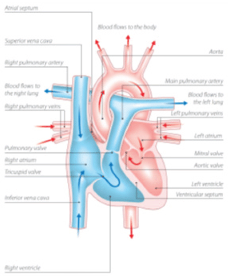
known as “Single Ventricle” is a congenital heart defect where both the left and the
right atrium opens into the left ventricle (Compare figure 1a. of normal heart and1b. of a heart with DILV). The right ventricle is either hypoplastic
or does not exist. It has a prevalence of 5-10 in 100,000 new-borns 3. DILV comprises about 1% of all congenital heart disease (CHD) 4. Median survival
of surgically uncorrected patients is about 14 years 5. Eisenmenger’s syndrome (ES, Eisenmenger’s reaction or tardive cyanosis) is a process
by which the left-to-right shunt caused by a congenital heart defect in a foetus causes
an increased flow through the pulmonary vasculature causing pulmonary hypertension
6 which over time causes increased pressure in the right side of the heart and reverses
the shunt into a right-to-left shunt. An informed consent was taken from the patient
for the purposes of this case report.
Figure 1a.
Normal Heart; 1. Understanding your child’s heart - Double inlet ventricle (British
Heart Association. 1st ed. 2016. Cited 28 November 2016. Available from: https://www.bhf.org.uk/publications/children-and-young-people/understanding-your-childs-heart---double-inlet-ventricle) Reprinted with permission from British Heart Association
The Case
The patient is a 29-year-old male from India, born in rural Kerala, came to an NGO
(primary level health care center, with free consultation) with chief complaints of
breathlessness on exertion since many years and increased discoloration of fingers,
tongue and limbs since the past 3-4 months.
- Breathlessness: Grade two (New York Heart Association classification) – Slight limitation of physical
activity. Ordinary physical activity results in fatigue, palpitation, dyspnoea. Patient
can walk for 0.5Km before the onset of dyspnoea. It is exaggerated on sustained physical
activity and is relieved on rest. There was a progressive increase in breathlessness
from the time of his birth till the patient was 10 years of age afterwards his symptoms
have improved and currently shows no progressive increase.
- Bluish discoloration of tips of fingers, tongue, lips: It is present at all times. It increases on exposure to cold climates, exposure to
cold water and other cold substances. This has been present since his childhood and
is temporarily improved with phlebotomy.
- Itching: Started five years back. Gradual in onset. Non-progressive, continuous. Increases
on taking a warm shower and relieved only after a phlebotomy.
No history of orthopnoea, paroxysmal nocturnal dyspnoea and platyopnoea, chest pain,
palpitation, syncope nor edema.
He gives a history of a single episode of hemoptysis three years back which has not
recurred since. Patient suffered from dengue when he was 18 years old and during laboratory
investigation for the same he was found to have high hemoglobin levels and has since
been asked to perform regular phlebotomies if his hemoglobin crossed 16g/dl (last
phlebotomy in 2015). He gives history of difficulty gaining weight as a child and
also history of repeated respiratory tract infections. There is no history of repeated
throat infections, diabetes mellitus, hypertension, thyrotoxicosis or bronchial asthma.
General examination revealed patient is moderately built and nourished (BMI: 18.51).
Patient has red conjunctiva; clubbing (Grade IV); cyanosis of lips, fingers and tongue.
His vital showed pulse: 92 beats per min, regular rhythm, normal in volume and character;
Respiratory rate: 26 breaths/min, abdomino-thoracic respiration. Jugular venous pulse
(JVP) was not raised.
Systemic examination
Cardiovascular System: Inspection (abnormal finding): Apex beat is visible in anterior axillary line in
the 6th intercostal space. There are no dilated veins, scars or sinuses. Palpation: Position of apex beat is confirmed and is of heaving type. Percussion: Indicates an enlarged heart. Right border of the heart being percussed in right parasternal
area. The upper border of the heart in the 3rd intercostal space in the parasternal
line. The left border of the heart in the 4th intercostal space. Auscultation: S1 and a loud S2 heard. Pan systolic murmur heard at the apex. Loud p2. Examination
of other systems reveals no abnormalities.
At this stage a provisional diagnoses of congenital cyanotic heart disease was made
with the possible differential diagnosis being double inlet ventricle; Tetralogy of
Fallot; patent foramen ovale; atrial septal defects; atrio-ventricular septal defects;
ventricular septal defects, and the persistent arterial duct.
Investigations
A review of the patient’s files the patient showed that the diagnosis of double inlet
left ventricle was made at a tertiary level hospital but no therapeutic interventions
were performed nor regular follow ups were made. Patient explained that financial
difficulties, lack of awareness for the need for follow up and absence of symptoms
that severely affected daily life were why he and his family did not feel the need
for regular follow up. Additional exams revealed:
- Complete blood count: Hemoglobin: 16.8 gm/dl; PCV:67.80 %; RBC:10.62 million/cu.mm
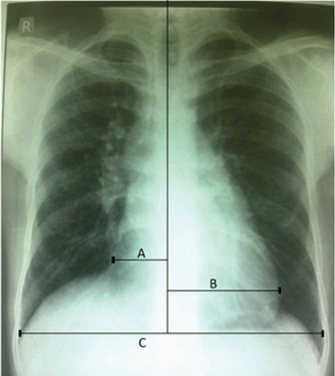
- Chest Radiography (Figure 2): Cardiomegaly.
- ECG: Sinus tachycardia, bi-atrial enlargement; left ventricular hyper trophy; probable
right ventricular hypertrophy
- ECHO: Left ventricle: Dominant; Right ventricle: Left outset; Great arteries are L-Malposed
with Aorta to the anterior and left from RV; Pulmonary artery posterior and right
from LV; Interventricular septum: Nonrestrictive bulboventricular foramen; Aorta arch:
Left sided.
Figure 1b.
Heart with Double Inlet Left Ventricle; Understanding your child’s heart - Double
inlet ventricle (British Heart association. 1st ed. 2016, cited 28 November 2016.
Available from: https://www.bhf.org.uk/publications/children-and-young-people/understanding-your-childs-heart---double-inlet-ventricle). Reprinted with permission from British Heart Association
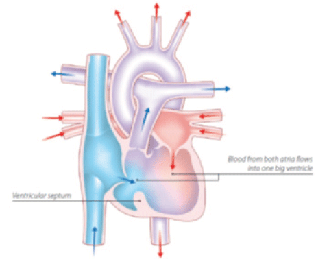
Figure 2.
X-ray Chest Posteroanterior View Showing Cardiomegaly [(A+B)/C = 0.54, A= 3.28cm,
B = 6.83cm, C = 18.52cm]
A final diagnosis of congenital heart disease; double inlet left ventrice, L-malposed
vessels without pulmonary stenosis, Eisenmenger Syndrome and absolute erythrocytosis
was made.
Management
The center at which the patient presented was not equipped neither with facilities
to treat a cardiac case nor a cardiologist for consultation on further management
strategies. The patient was counseled about the need for routine follow up treatment
with a single doctor and was advice to visit a tertiary care center. Up to this day
of writing this case report the patient has not visited a tertiary center and continues
with occasional phlebotomies.
Discussion
Etiology
DILV to be genetically determined by multiple genes. Recurrence & transmission risks
remain far below than that expected from medelian inheritance 7. In the polygenic model, the phenotype is presumed to result from additive effects
of multiple genes, interactions with other genes and environmental factors, and stochastic
effects 8.
Pathophysiology
In a heart with DILV blood from both the atria flow into the left ventricles from
here blood flows into the pulmonary circulation through the pulmonary artery and into
the systemic circulation by shunting (left to right shunt) through the bulboventricular
foramen and then entering the aorta (Figure 1b). The ratio of how much blood enters each circulation depends on the ratio of vascular
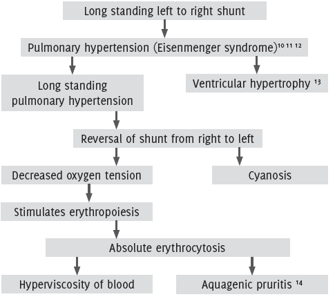
resistances in the two vascular beds 8. This results in a left to right shunt and later on its sequelae (Figure 3).
Figure 3.
Sequelae of double inlet left ventricle.
Prognosis
The actuarial survival rate without definitive repair was 57% at 1 year, 43% at 5
years, and 42% at 10 years for DILV. Moodie et al reported that 70% with well-formed
single left ventricles died before age 16, with an annual attrition rate of 4.8% 15. Usual causes of death are congenital heart disease, arrhythmias and sudden death
from unknown causes. A10-year mortality rate among untreated patients approached 30-40%
16. Common cause of death in these patients are hypoxemia and arrhythmia. They can also
die from congestive cardiac failure, thromboembolism and massive hemoptysis.
Conclusion
Unlike high-income countries where most congenital heart diseases are detected and
dealt with at birth low and middle-income nations often have to deal with cases that
present much later and should often be included in the differential diagnosis.
In India there are far too many patients and too few doctors (Sudhir Anand, Victoria
Fan. The health workforce in India, human resources for health observer series No.16.
World Health Organization; 2016. Available from: http://www.who.int/hrh/resources/16058health_workforce_India.pdf. Accessed June 8, 2017. The World Bank. World development indicators: Health systems.
Available from: http://wdi.worldbank.org/table/2.12#. Accessed June 8, 2017). Public health systems are overcrowded and private health
care is expensive and is mostly set up in urban India as compared to rural areas.
There does not exist a system in place for patient follow up after treatment or to
ensure that a patient has followed up at a higher center. Most patients seek symptomatic
treatment and once their acute episode has been controlled will insist on discharge
despite incomplete treatment of the cause. If the doctor refuses symptomatic treatment
the patient will simply move on to another doctor that is willing to do so, hence
compromising the health system. Financial difficulties provide another major problem;
expensive treatment, investigation and drugs assure lack of adherence to treatment
or failure to visit a doctor until the patient is significantly crippled. The Government
should upgrade primary health centers and increase the doctor – patient ratio and
implement strategies for the effective utilization of the present doctors such as
increasing the prominence of primary health centers thereby decreasing the load on
tertiary centers, medicines and basic scans such as the echocardiogram should be more
affordable. The process for improvement of health care in India has been initiated
and will require many more years to reach a level that can be compared to high-income
countries.
Acknowledgments
Special acknowledgments go to the Dr. Ashoojit Anand, MD Community medicine and the
Consultant ACCEPT.
Conflict of Interest Statement & Funding
The author has no conflict of interest to disclose.
Author Contributions
Collection and design the work/idea, collect data/obtaining results, analysis and
interpretation of data, write the manuscript, critical revision of the manuscript,
approval of the final version: RRA.
References
1. Restaino G, Dirksen MS, de Roos A. Long-term survival in a case of unoperated single ventricle. Int J Cardiovasc Imaging 2004; 20: 221–5.
2. Koito H, Ohkubo N, Suzuki J, Iwasaka T, Inada M. Prolonged survival in a patient with a single ventricle without pulmonary stenosis. Chest 1994; 106: 971–2. (Review)
3. Baldwin HS, Dees E. Embryology and physiology of the cardiovascular system. In: Gleason CA, Devaskar S, eds. Avery’s Diseases of the Newborn. 9th ed. Philadelphia, Pa: Saunders Elsevier; 2011:chap 50.
4. Franklin RC, Spiegelhalter DJ, Anderson RH, Macartney FJ, Rossi Filho RI, Douglas JM et al. Double-inlet ventricle presenting in infancy: I: Survival without definitive repair. J Thorac Cardiovasc Surg. 1991;101: 767–776.
5. Moodie DS, Ritter DG, Tajik AJ, O’Fallon WM. Long-term follow-up in the unoperated univentricular heart. Am J Cardiol 1984; 53: 1124–8.
6. Jensen AS, Iversen K, Vejlstrup NG, Hansen PB, Søndergaard L (April 2009). “[Eisenmenger syndrome]”. Ugeskrift for Laeger (in Danish) 171 (15): 1270–5.
7. Weigel TJ, Driscoll DJ, Michels VV. Occurrence of congenital heart defects in siblings of patients with univentricular
heart and tricuspid atresia. Am J Cardiol. 1989; 64: 768–771.
8. Burn J, Brennan P, Little J, Holloway S, Coffey R, Somerville J, Dennis NR, Allan L, Arnold R, Deanfield JE, Godman M, Houston A, Keeton B, Oakley C, Scott O, Silove E, Wilkinson J, Pembrey M, Hunter AS. Recurrence risks in offspring of adults with major heart defects: results from first
cohort of British collaborative study. Lancet. 1998; 351: 311–316.
9. Nelson DP, Schwartz SM, Chang AC. Neonatal physiology of the functionally univentricular heart. Cardiol Young. 2004; 14 (Suppl 1): 52–60.
10. Wood P. The Eisenmenger syndrome or pulmonary hypertension with reversed central shunt. Br Med J. Sep 27 1958;2(5099):755–62.
11. Vongpatanasin W, Brickner ME, Hillis LD, Lange RA. The Eisenmenger syndrome in adults. Ann Intern Med. May 1 1998;128(9):745–55
12. Diller GP, Gatzoulis MA. Pulmonary vascular disease in adults with congenital heart disease. Circulation. Feb 27 2007;115(8):1039–50.
13. Mohan H. Textbook of pathology. 6th ed. New Delhi: Jaypee Brothers Medical Pub; 2010:Chap 16, pg 421.
14. Abdel Naser MB, Gollnick H, Orfanos CE. Aquagenic pruritus as a presenting symptom of polycythemia vera. Dermatology. 1993;187:130–3.
15. Moodie DS, Ritter DG, Tajik AJ, O’Fallon WM. Long-term follow-up in the unoperated univentricular heart. Am J Cardiol.1984; 53: 1124–1128
16. Diller GP, Kempny A, Inuzuka R, Radke R, Wort SJ, Baumgartner H et al. Survival prospects of treatment naïve patients with Eisenmenger: a systematic review
of the literature and report of own experience. Heart. Sep 2014;100(17):1366–72.
Rahul Regi Abraham, 1 Dr. B. R. Ambedkar Medical College, Bengaluru, India.
2 Rajiv Gandhi University, India.
Editor: Huy Ming Lim
About the Author: Rahul Regi Abraham is a internal in Dr. B. R. Ambedkar Medical College, and the Rajiv
Gandhi University, in India.
Correspondence Rahul Regi Abraham, Email: abraham.rahul@gmail.com
Cite as: Abraham RR. Double Inlet Left Ventricle with Eisenmenger Syndrome in an Adult – A Case Report. Int J Med Students. 2017 Jan-Apr;5(1):53-56.
Copyright © 2017 Rahul Regi Abraham
International Journal of Medical Students, VOLUME 5, NUMBER 1, April 2017