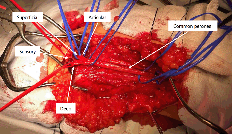
Intraoperative Picture of the Common Peroneal Nerve (CPN) with its Terminal and Side Branches after External Neurolysis, Decompression, and Complete Nerve Deliberation.
Highlights:Aleksa Mićić1, Stefan Radojević1, Lukas Rasulić2
doi: http://dx.doi.org/10.5195/ijms.2023.1956
Volume 11, Number 2: 139-143
Received 31 12 2022; Rev-request 25 01 2023; Rev-recd 07 03 2023; Accepted 08 03 2023
ABSTRACT
Background:A common peroneal nerve (CPN) injury located at the knee level, occurring as a consequence of hip surgery is described in the literature. However, there are only a few papers focusing on their surgical management, while there are no thoroughly analyzed cases following open reduction and internal fixation (ORIF) of the acetabular fracture. This paper aimed to describe such a case and discuss current trends in the surgical management of these patients.
Case:A 32-year-old woman was admitted to our department due to left-sided CPN palsy. The patient was injured in a traffic accident eight months earlier, followed by left hip dislocation and acetabular fracture. Following the acetabular fracture ORIF, a CPN palsy developed. The electromyoneurography (EMNG) and ultrasound (US) indicated a nerve lesion at the knee level. The surgical treatment included external neurolysis, decompression, and complete nerve deliberation, with the preservation of all nerve branches. The patient reported immediate relief and completely recovered 8 months following the surgery (Medical Research Council (MRC) grade = 5, Visual Analogous Scale (VAS) = 0).
Conclusion:The cause of CPN palsy following hip surgery may not always be located in the hip region. A detailed anamnesis, physical examination, and diagnostic evaluation are necessary for the proper surgical management of these patients. In addition to the EMNG, the US should be essential in preoperative planning and choosing the most effective surgical strategy.
Keywords: Orthopedic Procedures; Common Peroneal Nerve Entrapment; Neurosurgery; Hip fractures (Source: MeSH-NLM).
Peroneal nerve dysfunction often referred to as common peroneal nerve (CPN) palsy presents a functional deficit, characterized by an inability to dorsiflex the foot (foot drop) with consequential disability and a significant decrease in patients' quality of life.1-3 The cause of dysfunction may lie in central nerve structures, spinal nerve roots, sacral plexus, sciatic nerve (SN), the CPN, or its deep branch.4
The incidence of CPN palsy following the most common procedure in hip surgery: the total hip arthroplasty (THA), ranged up to 8%, depending on the inclusion and exclusion criteria of the reviewed studies.5-7 In most cases, the cause of the palsy involves SN injury at the hip region and may be a result of various etiologies, such as direct nerve injury, excessive nerve stretch, postoperative hematoma, or infection.7 Rarely, the palsy may develop as a consequence of CPN injury at the knee level, and there are only a few papers concerning their surgical management, 8, 9 with a recently reported incidence of 0.48%.10
The open reduction and internal fixation (ORIF) of the acetabular fracture is performed less often than the THA, thus the associated CPN injuries at the knee level are extremely rare.11, 12 To the best of our knowledge, a thoroughly analyzed case of surgical treatment of such injury is not reported in the English medical literature. This paper aimed to describe a such case and discuss current trends in the surgical management of these patients.
A 32-year-old woman was admitted to our department due to left-sided CPN palsy. The patient was injured in a traffic accident eight months earlier, followed by left hip dislocation and acetabular fracture. After admission to the emergency care unit, the hip dislocation was repositioned. Two weeks later, ORIF of the acetabular fracture was performed, resulting in ipsilateral CPN palsy.
Upon admission to our department, clinical findings included left-sided incomplete CPN palsy (Medical Research Council (MRC) grade 2), pain in the lateral lower leg (Visual Analogue Scale (VAS) score 3), and gait disturbances. Using electromyoneurography (EMNG), the peroneal nerve lesion was located at the knee level. Reduced nerve conduction was noted in the tibialis anterior (TA) and extensor digitorum brevis (EDB) muscles, while there were no changes in the short head of the biceps femoris (shBF) and muscles innervated by the tibial nerve. The ultrasound (US) findings indicated a suspectable CPN compression due to visible nerve thickening proximal to the fibular tunnel. The PNSQoL and SF-36 scores indicated a significant decline in the patient's quality of life.
Following GETA, the external neurolysis, decompression, and complete nerve deliberation were performed through the popliteal approach, with the preservation of all nerve branches (Figure 1). The nerve was thinned at the site of the previous compression. There were no signs of nerve bruising.
Figure 1.Intraoperative Picture of the Common Peroneal Nerve (CPN) with its Terminal and Side Branches after External Neurolysis, Decompression, and Complete Nerve Deliberation.
The patient reported immediate relief following the surgery. 2 months after the surgery, there were signs of motor recovery, with the improvement of foot dorsiflexion and gait performance. 3 months after the surgery, a significant motor recovery was noted with insignificant gait disturbances mostly due to pain. A complete motor and sensory recovery was achieved 8 months following the surgery (MRC = 5, VAS = 0). In order to assess the patient's postoperative quality of life, a prolonged follow-up is needed.
The etiology of nerve injury in patients with CPN palsy is various. Regarding the CPN lesions at the knee level, the traumatic causes may include direct nerve damage, or indirect during orthopedic trauma such as hip or knee dislocation, or fibula fracture.13, 14 The iatrogenic causes may include surgical interventions in the lower extremity, improper positioning during general anesthesia, or application of the splints, casts, wrappings, and bandages.14, 15 The idiopathic causes may include increased anatomic risk factors and consequential CPN entrapment at the fibular tunnel,16 or formation of an intraneural ganglion cyst,17, 18 while neoplastic etiology mostly include nerve schwannomas.19
Surgical treatment of the CPN palsy following trauma and associated orthopedic interventions must be planned well, due to the various mechanisms of injury and different locations at which the nerve may be damaged. Contrary to compression injuries that are easily managed by a simple surgical decompression, injuries due to traction or contusion may require more complex procedures such as tendon transfer or nerve repair.3, 20, 21
Knowing the exact time of the nerve injury is important in determining the most effective surgical strategy, in terms of choosing the most appropriate procedure and setting the timing for surgery.3, 22, 23 While surgical decompression is indicated after 3 months of conservative management without the signs of motor recovery, the nerve repair should not be performed later than 12 months post-injury.24 On the other hand, tendon transfers are reserved for cases with poor recovery capacity due to extensive nerve injury or due to exceeding the timing for nerve repair.3
Based on our experience,3, 22, 23 in some cases it is difficult to determine if the injury is acquired during the trauma or during the orthopedic intervention because the patient is immobilized and unaware of the present deficit. Regarding the patient described in this paper, the hip dislocation and its repositioning could have been a cause of the palsy due to the traction and contusion of the SN.25 However, this was not the case. During the period between the trauma and the performed ORIF, both the patient and the medical staff were aware of the preserved CPN function and reported the palsy immediately following the intervention.
We could not provide the surgery for the patient when it was indicated because she had been recovering for a long time after the trauma and came to our clinic for evaluation 7 months later. Although, it seems that the timing was not a factor that affected the outcome of our patient, which is in accordance with a similar case that occurred following the THA.8 In their paper, the authors performed CPN decompression 8 months following the injury and achieved almost complete motor recovery. A poorer recovery in their patient compared to ours could be a consequence of a more serious CPN damage or a different decompression procedure that was used.
Similar to the study by Wilson et al,9 we used the EMNG to detect a reduced CPN conduction and locate the nerve lesion at the knee level. However, in their study, 22% of the patients had no motor unit potentials (MUPs) in the short head of the biceps femoris muscle (BFsh), indicating that the lesion may have extended proximally in relation to the fibular tunnel.26 Even though there was a tendency for better outcomes in those who had the BFsh MUPs, the CPN decompression had positive effects on motor recovery in some of the cases with reduced BFsh MUPs.9 Regarding our patient, there was no reduction in the BFsh MUPs, and complete motor recovery was achieved. Therefore, a simultaneous CPN and SN injury should be considered in cases that fail to recover.27
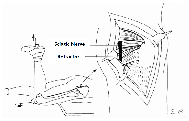
Compared to EMNG, the US is more precise in determining the exact location of the nerve injury, while in comparison to magnetic resonance imaging (MRI), it has a higher sensitivity in detecting peripheral nerve pathology and understanding the mechanism of injury.27 Thus, the US plays a very important role in diagnosing and managing the patients with peripheral nerve injuries. It was already discussed in the literature that a CPN palsy following the acetabular fracture ORIF,11 usually performed in the Cocker-Lagenback position (Figure 2), may be a consequence of SN traction by the retractors and consequential CPN compression due to its reduced mobility at the fibular tunnel. This mechanism may be applied to our patient, considering the US finding that revealed nerve thickening proximal to the fibular tunnel and intraoperative finding of the nerve (Figure 1) which was thinned at the site of the previous compression.
Figure 2.Drawing to Present the Proximity of the Sciatic nerve to the Retractors during the Acetabular Fracture ORIF Performed in the Cocker-Lagenback Position, with Courtesy of Strahinja Gligorijević, BA (Faculty of Biology, University of Belgrade). Flexion of the Knee is a Measure for preventing the associated CPN injury.
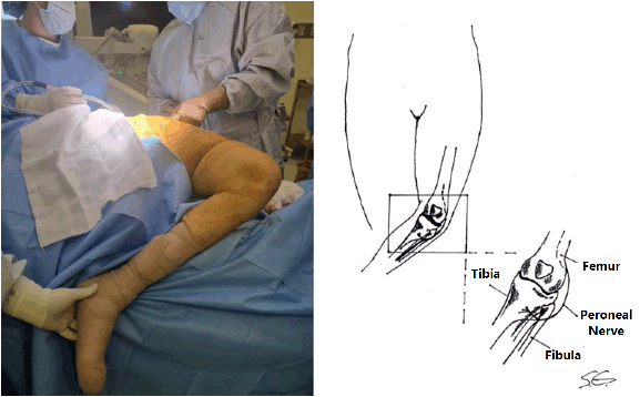
Compared to the aforementioned mechanism of CPN injury following the acetabular fracture ORIF, there are some differences in cases that occur following THA. In the posterior approach for THA,28 the SN is often visualized, and its traction may result in CPN compression at the fibular tunnel. However, in THA procedures in which the SN is not visualized,29 such as the Smith Petersen and Watson Jones approach, the cause of the CPN injury is probably SN stretching due to leg manipulation (Figure 3). Compared to the SN stretching by the retractors, in cases with leg manipulation, the length of the nerve stretch may be longer, resulting in more serious CPN entrapment or even a traction injury. In their study,9 Wilson et al. did not use the US to confirm isolated CPN entrapment and to differentiate cases with traction injury. This may be the factor influencing poorer postoperative recovery in some of their patients and indicates the importance of US usage in the preoperative evaluation of such cases.
Figure 3.Leg Manipulation during the Antero-Lateral Approaches for Total Hip Arthroplasty (THA): (A) Intraoperative picture, with courtesy of dr Norma Izchel Orozco Aponte (Clinica de Nervio Periférico en Puebla, Mexico); (B) Drawing with Courtesy of Strahinja Gligorijević, BA (Faculty of Biology, University of Belgrade, Serbia).
Slabost peronealnog živca manifestuje se padom stopala, funkcionalnim deficitom koji značajno remeti kvalitet života pacijenta. Lokalizacija nervnog oštećenja, koje uzrokuje pad stopala, može biti na relaciji od mozga, preko kičmene moždine do nerava. Povreda peronealnog živca na nivou kolena, kao posledica operacije kuka je već opisana u literature. Međutim, objavljeno je samo nekolicina radova koji se bave hirurškim lečenjem ovakvih pacijenata, dok nema detaljno analiziranih slučajeva nakon otvorene redukcije i unutrašnje fiksacije karličnog preloma. Cilj ovog rada bio je da se opiše jedan takav slučaj, kao i da se prodiskutuju savremeni pristupi u lečenju ovih pacijenata.
Tridesetdvogodišnja žena je upućena na Kliniku za neurohirurgiju Univerzitetskog kliničkog centra Srbije, zbog levostranog pada stopala i bola u potkolenici. Povređena je u saobraćajnoj nesreći 8 meseci ranije, kada je zadobila dislokaciju kuka i frakturu acetabulum-a na levoj strani. Pad stopala nastupio je nakon otvorene redukcije i interne fiksacije preloma. Pomoću ultrazvuka i elektromioneurografije, povreda nerva locirana je na nivou kolena. Načinjena je dekompresija, deliberacija i spoljašnja neuroliza n. peroneus – a, kroz poplitealni pristup. Olakšanje u vidu smanjenja bola je prijavljeno odmah nakon operacije, a kompletni oporavak je postignut 8 meseci kasnije.
Uzrok pada stopala nakon operacije kuka ne mora uvek biti na nivou kolena. Detaljna anamneza, fizikalni pregled i dijagnostička evaluacija su neophodni za pravilno lečenje ovih pacijenata. Uz elektromioneurografiju, ultrazvuk bi trebalo da bude esencijalan u preoperativnom planiranju i odabiru najefektivnije hirurške strategije.
The authors would like to extend their sincere thanks to Andrija Savić MD, Ph.D. and Jovan Grujić MD (Clinic for Neurosurgery, University Clinical Centre of Serbia, Serbia), Norma Izchel Orozco Aponte MD (Clinica de Nervio Periférico en Puebla, Mexico), Jovan Vesic MD (Clinic for Orthopedic Surgery and Traumatology, University Clinical Centre of Serbia, Serbia), and Milan Lepić MD, Ph.D. (Clinic for Neurosurgery, Military Medical Academy) for continuous support and help during the writing of this article.
The Authors have no funding, financial relationships or conflicts of interest to disclose.
Conceptualization: AM, LR. Formal Analysis: AM, SR, LR. Investigation: AM, SR, LR. Resources: LR. Supervision: LR. Validation: LR. Writing - Original Draft: AM. Writing - Review Editing: AM.
1. Aprile I, Caliandro P, La Torre G, Tonali P, Foschini M, Mondelli M, et al. Multicenter study of peroneal mononeuropathy: clinical, neurophysiologic, and quality of life assessment. J Peripher Nerv Syst. 2005;10(3):259–68.
2. Poage C, Roth C, Scott B. Peroneal Nerve Palsy: Evaluation and Management. JAAOS - Journal of the American Academy of Orthopaedic Surgeons. 2016;24(1):1–10.
3. Rasulić L, Nikolić Ž, Lepić M, Savić A, Vitošević F, Novaković N, et al. Useful functional recovery and quality of life after surgical treatment of peroneal nerve injuries. Front Surg. 2022;9:1005483.
4. Carolus AE, Becker M, Cuny J, Smektala R, Schmieder K, Brenke C. The Interdisciplinary Management of Foot Drop. Dtsch Arztebl Int. 2019;116(20):347–54.
5. Park JH, Hozack B, Kim P, Norton R, Mandel S, Restrepo C, et al. Common peroneal nerve palsy following total hip arthroplasty: prognostic factors for recovery. J Bone Joint Surg Am. 2013;95(9):e55.
6. Zappe B, Glauser PM, Majewski M, Stöckli HR, Ochsner PE. Long-term prognosis of nerve palsy after total hip arthroplasty: results of two-year-follow-ups and long-term results after a mean time of 8 years. Archives of Orthopaedic and Trauma Surgery. 2014;134(10):1477–82.
7. Hasija R, Kelly JJ, Shah NV, Newman JM, Chan JJ, Robinson J, et al. Nerve injuries associated with total hip arthroplasty. J Clin Orthop Trauma. 2018;9(1):81–6.
8. Makhdom AM. Common Peroneal Nerve Palsy at the Level of Proximal Fibula After Total Hip Arthroplasty: A Case Report. Cureus. 2022;14(10):e30741.
9. Wilson TJ, Kleiber GM, Nunley RM, Mackinnon SE, Spinner RJ. Distal peroneal nerve decompression after sciatic nerve injury secondary to total hip arthroplasty. J Neurosurg. 2018;130(1):179–83.
10. Georgeanu VA, Russu OM, Obada B, Iliescu MG, Popescu MN, Iliescu DM, et al. Common peroneal nerve palsy after primary total hip arthroplasty. Int Orthop. 2022;46(9):1963–70.
11. Simske NM, Krebs JC, Heimke IM, Scarcella NR, Vallier HA. Nerve Injury With Acetabulum Fractures: Incidence and Factors Affecting Recovery. J Orthop Trauma. 2019;33(12):628–34.
12. Ramanan M, Chandran KN. Common peroneal nerve decompression. ANZ J Surg. 2011;81(10):707–12.
13. Seidel JA, Koenig R, Antoniadis G, Richter HP, Kretschmer T. Surgical treatment of traumatic peroneal nerve lesions. Neurosurgery. 2008;62(3):664–73; discussion -73.
14. Lezak B, Massel DH, Varacallo M. Peroneal Nerve Injury. StatPearls. Treasure Island (FL): StatPearls Publishing. Copyright © 2023, StatPearls Publishing LLC.; 2023.
15. Antoniadis G, Kretschmer T, Pedro MT, König RW, Heinen CP, Richter HP. Iatrogenic nerve injuries: prevalence, diagnosis and treatment. Dtsch Arztebl Int. 2014;111(16):273–9.
16. Fortier LM, Markel M, Thomas BG, Sherman WF, Thomas BH, Kaye AD. An Update on Peroneal Nerve Entrapment and Neuropathy. Orthop Rev (Pavia). 2021;13(2):24937.
17. Kim D, Choi JG, Son BC. Peroneal Nerve Palsy Due to Subparaneurial Ganglion Cyst, a Rare Variant of Intraneural Ganglion Cyst. Asian J Neurosurg. 2018;13(4):1225–8.
18. Bucher F, Maerz V, Obed D, Vogt PM, Weyand B. Intraneural Ganglion of the Peroneal Nerve-A Rare Cause of Pediatric Peroneal Nerve Palsy: A Case Report. European J Pediatr Surg Rep. 2022;10(1):e33–e6.
19. Milenković SS, Mitković MM. Common peroneal nerve schwannoma. Hippokratia. 2018;22(2):91.
20. Humphreys DB, Novak CB, Mackinnon SE. Patient outcome after common peroneal nerve decompression. J Neurosurg. 2007;107(2):314–8.
21. Emamhadi M, Bakhshayesh B, Andalib S. Surgical outcome of foot drop caused by common peroneal nerve injuries; is the glass half full or half empty? Acta Neurochir (Wien). 2016;158(6):1133–8.
22. Rasulić L, Djurašković S, Lakićević N, Lepić M, Savić A, Grujić J, et al. Surgical Treatment of Radial Nerve Injuries Associated With Humeral Shaft Fracture—A Single Center Experience. Frontiers in Surgery. 2021;8.
23. Rasulić L, Đjurašković S, Lakićević N, Lepić M, Savić A, Grujić J, et al. Etiological and epidemiological characteristics of surgically treated radial nerve lesions: A 20-year single-center experience. Frontiers in Surgery. 2022;9.
24. George SC, Boyce DE. An evidence-based structured review to assess the results of common peroneal nerve repair. Plast Reconstr Surg. 2014;134(2):302e–11e.
25. Cornwall R, Radomisli TE. Nerve Injury in Traumatic Dislocation of the Hip. Clinical Orthopaedics and Related Research®. 2000;377:84–91.
26. Thatte H, De Jesus O. Electrodiagnostic Evaluation Of Peroneal Neuropathy. StatPearls. Treasure Island (FL): StatPearls Publishing. Copyright © 2022, StatPearls Publishing LLC.; 2022.
27. Wijntjes J, Borchert A, van Alfen N. Nerve Ultrasound in Traumatic and Iatrogenic Peripheral Nerve Injury. Diagnostics (Basel). 2020;11(1).
28. Angerame MR, Dennis DA. Surgical approaches for total hip arthroplasty. Annals of Joint. 2018;3(5).
29. Weale AE, Newman P, Ferguson IT, Bannister GC. Nerve injury after posterior and direct lateral approaches for hip replacement. A clinical and electrophysiological study. J Bone Joint Surg Br. 1996;78(6):899–902.
Aleksa Mićić, 1 MD up to 3 months after graduation/Clinic for Neurosurgery, University Clinical Centre of Serbia, Belgrade Serbia.
Stefan Radojević, 1 MD up to 3 months after graduation/Clinic for Neurosurgery, University Clinical Centre of Serbia, Belgrade Serbia.
Lukas Rasulić, 2 MD, Ph.D., Full Professor/Faculty of Medicine, University of Belgrade, Belgrade Serbia/Clinic for Neurosurgery, University Clinical Centre of Serbia, Belgrade, Serbia.
About the Author: Aleksa Mićić is MD up to 3 months after graduation, currently engaged as an intern at the University Clinical Centre of Serbia. He has 2 years of experience as an undergraduate research fellow at the Department for Peripheral Nerve Surgery, Functional Neurosurgery, and Pain Management Surgery, Clinic for Neurosurgery, University Clinical Centre of Serbia, and 7 years of experience as an associate at Petnica Science Center.
Correspondence: Aleksa Mićić. Address: Pasterova 2, Beograd 11000, Serbia. Email: aleksamicic.md@gmail.com
Editor: Francisco J. Bonilla-Escobar; Student Editors: Eugenia M. Ramos-Dávila & Hang-Long (Ron) Li; Proofreader: Laeeqa Manji; Layout Editor: Ana Maria Morales; Process: Peer-reviewed
Cite as Mićić A, Radojević S, Rasulić L. Peroneal Nerve Injury due to Hip Surgery Located at the Knee Level: A Case Report. Int J Med Stud. 2023 Apr-Jun;11(2):139-43.
Copyright © 2023 Aleksa Mićić, Stefan Radojević, Lukas Rasulić
This work is licensed under a Creative Commons Attribution 4.0 International License.
International Journal of Medical Students, VOLUME 11, NUMBER 2, April 2023