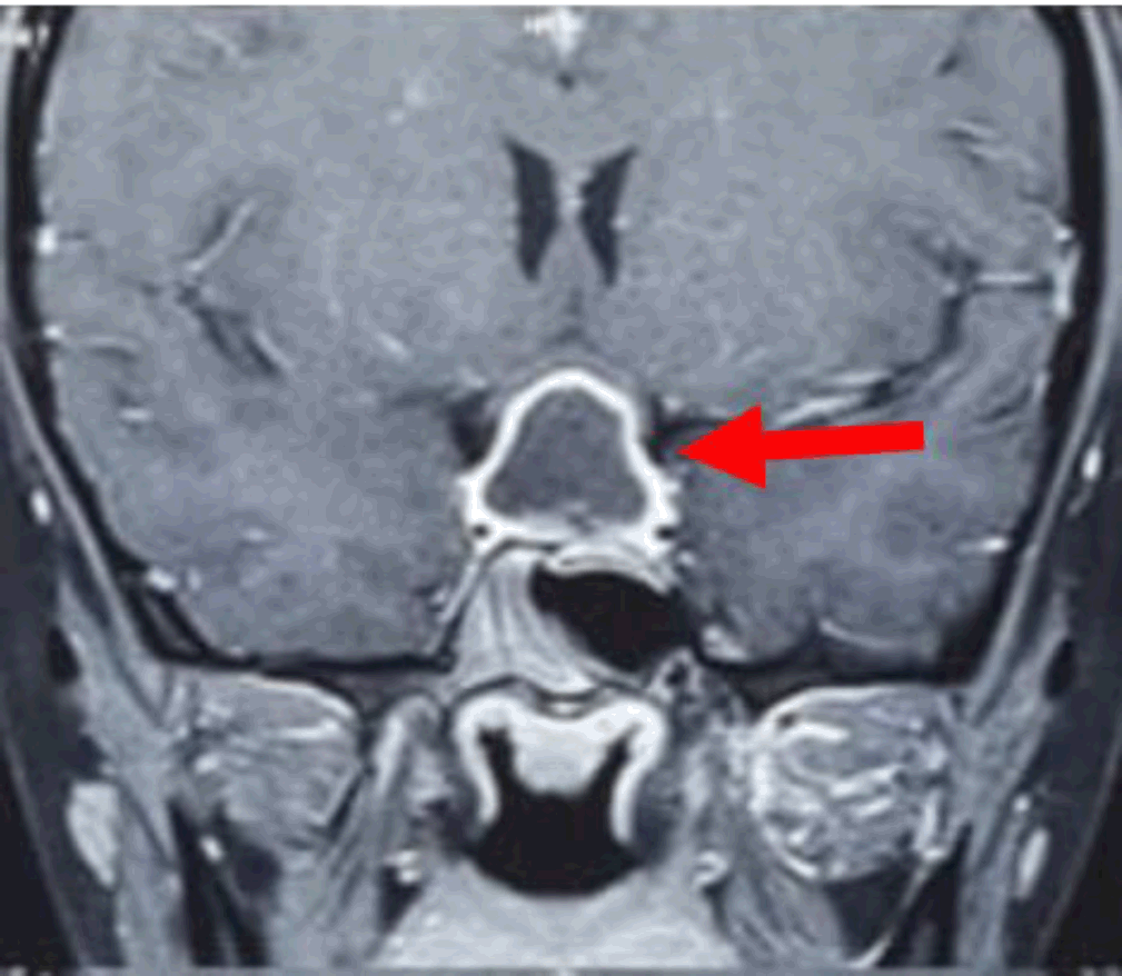
MRI Brain Coronal Study Showing Sellar Mass with Suprasellar Extension Causing Optic Chiasm Compression.

Highlights:Arwa Moiz Jamali1, Rakeshkumar Luhana2
doi: http://dx.doi.org/ijms.2024.2128
Volume 12, Number 3: 334-337
Received 25 06 2023; Rev-request 05 08 2023; Rev-request 06 05 2024; Rev-recd 21 08 2023; Rev-recd 20 05 2024; Accepted 12 09 2024
ABSTRACT
Background:Tuberculosis of the central nervous system is an uncommon but one of the most severe forms. It manifests as tuberculoma and tuberculous meningitis, with the majority of cases affecting children and immunocompromised patients. Overall, tuberculomas make up to 0.15–2% of all intracranial lesions but sellar tuberculoma is extremely rare.
The Case:An 18-year-old female patient presented with complaint of generalized weakness, eye pain and headache for 3–4 months. Magnetic resonance imaging (MRI) of brain showed sellar and suprasellar space occupying lesion. Trans sphenoidal approach was used to remove the lesion completely. A sellar tuberculoma was confirmed on pathological evaluation and the patient was put on postoperative anti-tubercular therapy.
Conclusion:Although rare, intracranial tuberculomas, particularly those that originate in the sellar, are notorious for mimicking pituitary tumors by jeopardizing pituitary hormonal function and applying compressive forces on surrounding intracranial structures. However, a prompt assessment can help overcome this diagnostic difficulty with the timely initiation of anti-tubercular therapy (ATT).
Worldwide tuberculosis (TB) is amongst the most lethal infectious diseases. In 2021, it was predicted that 10.6 million people worldwide would contract TB. More than two thirds of all cases of TB worldwide were found in eight countries highest being in India (28%).1 Although TB is primarily a respiratory tract disease, 1% of cases also impact the central nervous system (CNS). These cases are the most severe form of the disease and have the greatest rates of morbidity and mortality.2
The most prevalent form of CNS TB is tuberculous meningitis, however in <5% of cases, TB of the CNS can manifest as a granulomatous mass lesion known as a tuberculoma.3 Before the advent of chemotherapy, 30–50% of space occupying lesions (SOL) in both adults and children were tuberculomas.4 The prevalence of intracranial tuberculomas has decreased as a result of chemotherapeutic drugs and improvements in socioeconomic situations. However, they still account for 0.15–4% of intracranial lesions, and the majority of the affected are individuals from low- and middle-income nations.5,6
Intracranial tuberculomas are usually found in the cerebellum and cerebral cortex, however they have also been reported in the thalamus, cerebello-pontine angle, brainstem, basal ganglion, pineal region, optic pathways, in the ventricles, and the aqueduct. Of this isolated sellar tuberculoma is quite uncommon.
This type of tuberculoma is characterized by growth of tuberculous lesion affecting the sellar region of the brain which houses the pituitary gland. Its clinical importance lies in its potential to cause a range of neurological and endocrine symptoms due to its proximity to the pituitary gland and optic pathways.
As a result, understanding the presentation and diagnosis of sellar tuberculomas is crucial for providing timely and effective medical and surgical management. Given the limited existing literature on it, by delving into these aspects, this case report aims to contribute valuable insights that can enrich the existing knowledge base surrounding this rare manifestation, ultimately fostering a better understanding of its nature and optimal approaches for addressing it.
An 18-year-old female patient presented to outpatient department with chief complaints of headache, eye pain and generalized weakness with an onset of 3–4 months. She denied any history of fever, cough, weight loss, or anorexia. There was no history of any known TB exposure, no notable comorbid conditions or pertinent social factors. No previous history of medication use was noted except analgesics. On examination, the patient was alert and fully oriented to time, place, and person. Vital signs were stable, with a pulse rate of 82 beats per minute, blood pressure of 110/80 mmHg, oxygen saturation (SpO2) of 98%, and body temperature of 98.6°F. No neurological deficits or abnormalities in other systems were noted during examination. On investigations low levels of T3, T4, TSH were found (Table 1). Magnetic resonance imaging (MRI) revealed a sellar and suprasellar space-occupying lesion measuring 1.4 × 1.9 × 1.0 cm (Figure 1).
Table 1.Complete Laboratory Profile of an 18-Year-Old Female with Sellar Tuberculoma.
| Investigations | Patient Values | Normal Ranges |
|---|---|---|
| Hemoglobin | 12.40 g/dl | 12.1–15.1 g/dL (females) and 13.8–17.2 g/dL (males) |
| Total leucocyte count | 7860/µL | 4000 to 11000/microliter |
| Platelet count | 3.13 lakh/µL | 150,000 to 450,000/µL |
| Random Blood Sugar | 89 mg/dl | 70 to 140 mg/dL |
| Serum creatinine | 0.77 mg/dL. | 0.6 to 1.2 mg/dL. |
| T3 | 1.2 ng/mL | 0.8–2.0 ng/ml |
| T4 | 6 ng/dL | 0.8 ng/dL to 1.8 ng/dL |
| TSH | 0.13 microU/mL | 0.4 to 4.0 microU/mL |
| HIV, HCV, HBSAG | Negative |
MRI Brain Coronal Study Showing Sellar Mass with Suprasellar Extension Causing Optic Chiasm Compression.

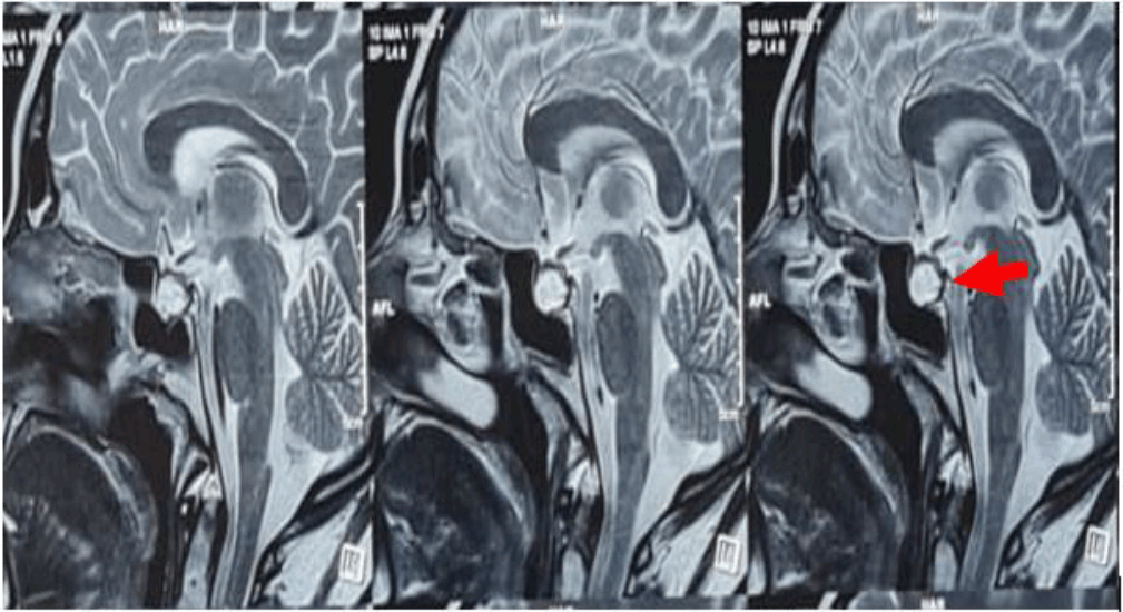
On assessing the hormone profile hypopituitarism was noted (Table 2). After proper preoperative workup, endoscopic transnasal transsphenoidal approach with sellar and suprasellar space occupying lesion excision and sellar floor reconstruction with fat graft under general anesthesia was carried out (Figure 2). Intra operative and post operative period was uneventful. Post operative strict input output, thyroid profile and sodium were monitored to evaluate the function of the pituitary hormones. As the lesion was surgically removed the pressure on surrounding structures, such as the optic nerves and pituitary gland diminished leading to reduced intensity and frequency of patient's headache and gradually its resolution. Eye pain and weakness also improved gradually, and patient was vitally stable. Histopathology of the lesion revealed caseating granuloma. Ziehl Neelsen staining demonstrated acid-fast bacilli most likely tuberculoma. The patient was prescribed rifampicin (450 mg), isoniazid (300 mg), ethambutol (800 mg), and pyrazinamide (750mg) for 1 year (3 months intensive phase and 9 months continuation phase) and also prednisolone and thyroxine to address the endocrinological deficits. Regular monitoring and administration of prednisolone and thyroxine stabilized hormone levels.
Table 2.Hormonal Profile of an 18-Year-Old Female with Sellar Tuberculoma.
| Hormone value | Patient Values | Normal Ranges |
|---|---|---|
| Serum cortisol | 0.4 μg/dL | 5 to 23 μg/dL |
| LH | 0.5 mIU/mL | Female (follicular phase): 1.9 to 12.5 mIU/mL |
| Female (mid-cycle surge): 8.7 to 76.3 mIU/mL | ||
| Female (luteal phase): 0.5 to 16.9 mIU/mL | ||
| FSH | 3.5 mIU/mL | Female (follicular phase): 3.5 to 12.5 mIU/mL |
| Female (mid-cycle surge): 4.7 to 21.5 mIU/mL | ||
| Female (luteal phase): 1.7 to 7.7 mIU/mL | ||
| Prolactin | 129 μg/dL | 2 to 29 μg/dL |
| GH | 0.0536 μg/dL | 0 to 10 μg/dL |
| IGF1 | 38.80 ng/mL | Age 0 to 18: 75 to 325 ng/mL |
MRI Showing Postoperative Sellar Floor Reconstruction with Fat Graft.

Pituitary adenomas are the most common lesions of the sellar region, but it is important to consider atypical non-adenohypophyseal lesions, including inflammatory and infectious conditions into account when making a sellar mass differential diagnosis. TB (tuberculoma) should be considered particularly in India. Sellar tuberculomas are fairly uncommon pathology.7 From 1924–2019, 106 cases could be retrieved from the literature worldwide, out of which 51 cases were reported from India.8
Tuberculomas usually occur as part of a systemic infection, following hematogenous spread; however isolated lesions have been reported. In our case there was no evidence of systemic or primary active TB based on the patient's history and appropriate investigations. Also, patient's medical history revealed no prior diagnosis of immune compromise like HIV or immunosuppressive medications which are potential risk factor for this condition. In patients, headache is the most common symptom occurring earlier and is accompanied by visual disturbances. Variable levels of anterior pituitary dysfunction can later arise with or without central diabetes insipidus due to their compressive effects. Endocrinological evaluation varies from hypopituitarism to hyperprolactinemia. Majority of the patients are young females with a mean age of 36 years.9
Pituitary tuberculoma looks radiographically similar to an adenoma, making a straightforward identification of the disease challenging, particularly in the absence of a pulmonary TB history. Pituitary tuberculoma is known to cause thickening of the stalk. However, this nonspecific finding can also be found in inflammatory conditions like eosinophilic granuloma, granulomatous hypophysitis and sarcoidosis.10 Additionally, the tuberculoma suprasellar extension renders a proper pituitary stalk evaluation on neuroimaging studies extremely challenging. Consequently, in order to make a precise diagnosis surgery is required and to decompress the optic chiasma. Trans-sphenoidal route is one the safest approach as it prevents cerebrospinal fluid contamination.7 Sub-frontal approach after craniotomy is an alternative.
Histopathological examination is required to make the final diagnosis. Tuberculomas are classically characterized by a central area of caseation necrosis surrounded by epithelioid macrophages, lymphocytes, plasma cells and Langhans giant cells. On Ziehl-Neelsen staining, acid fast bacilli may or may not be visible. Cerebrospinal fluid and specimens can also be used for the diagnosis using the polymerase chain reaction technique. Anti-tubercular therapy (ATT) is mandatory, and in cases where hypopituitarism symptoms and signs are present, hormone replacement therapy may need to be started.11 Our patient was prescribed ATT with 4-drug regimen of isoniazid, rifampin, pyrazinamide, ethambutol along with prednisolone and thyroxine. While it's prudent to obtain tissue diagnosis before initiating targeted treatment, in cases where pituitary tuberculosis is suspected, ATT can be commenced based on clinical suspicion alone. There are documented instances of significant reduction in the size of pituitary tuberculoma following initiation of ATT without confirmed tissue diagnosis.12 This underscores the potential efficacy of medical management as a viable alternative to surgical intervention, particularly in cases where surgery poses significant risks or is not feasible due to patient factors.
Regular follow-up care is essential to monitor and manage any persistent symptoms or complications. In cases where treatment is delayed or ineffective, permanent endocrine dysfunction can be a challenging long-term outcome and can significantly compromise a patient's quality of life.13 While this case report sheds light on sellar tuberculoma, its findings might lack generalizability and offer limited causality assessment due to uniqueness of individual cases. To address this, future studies could include case series and retrospective or prospective analyses to provide a more comprehensive understanding of the condition.
“My journey through treatment for a pituitary tuberculoma has been a rollercoaster of emotions. Planning my treatment involved discussions with my healthcare team, weighing the pros and cons of medications and potential surgery. The emotional impact was immense, as fear and uncertainty about the future consumed me. Support from my loved ones and healthcare professionals became my lifeline. I am quite satisfied with the treatment provided and my symptoms started to relieve few days after surgery. Coping with side effects, adjusting my lifestyle, and undergoing regular monitoring were challenging but necessary steps”.
We report a case of sellar tuberculoma, an uncommon lesion that should be considered when making a sellar lesion differential diagnosis. Additionally, we also stress upon the importance of histopathological examination for the final diagnosis and postoperative anti-tubercular therapy in management of such cases. Prompt evaluation, rapid diagnosis, appropriate surgical management and timely starting of the anti-tuberculous regimen in patients result in great outcomes and noticeably improved clinical symptoms. This case emphasizes the diagnostic challenge of pituitary tuberculoma and the need for further research on specific diagnostic tools and long-term treatment efficacy.
Venus Superspecialty Hospital, Vadodara, India
The Authors have no funding, financial relationships or conflicts of interest to disclose.
Conceptualization: A J. Data Curation : A J. Investigation: A J. Writing – Original Draft: A J. Visualization: A J. Project Administration: A J. Writing – Review & Editing: R L. Supervision: R L.
1. Global Tuberculosis Report 2022. Geneva: World Health Organization; 2022.
2. DeLance AR, Safaee M, Oh MC, Clark AJ, Kaur G, Sun MZ, et al Tuberculoma of the central nervous system. J Clin Neurosci. 2013;20(10):1333–41.
3. Ayele B, Wako A, Shewaye A, Tessema A. Sellar Tuberculoma: A rare presentation in a 30-year-old Ethiopian woman: case report. Afr J Neurol Sci. 2019;38(1):50–53.
4. Dastur HM. Tuberculoma. In: Vinken PJ, Bruyn GW, editors. Handbook of Clinical Neurology, vol. 18. New York: American Elsevier; 1975:413–26.
5. DeAngelis LM. Intracranial tuberculoma: case report and review of the literature. Neurology. 1981;31(9):1133–6.
6. Maurice-Williams RS. Tuberculomas of the brain in Britain. Postgrad Med J. 1972;48(565):679–81.
7. Ranjan A, Chandy MJ. Intrasellar tuberculoma. Br J Neurosurg. 1994;8(2):179–85.
8. Kumar T, Nigam JS, Jamal I, Jha VC. Primary pituitary tuberculosis. Autops Case Rep. 2020;11: e2020228.
9. Furtado SV, Venkatesh PK, Ghosal N, Hegde AS. Isolated sellar tuberculoma presenting with panhypopituitarism: clinical, diagnostic considerations and literature review. Neurol Sci. 2011;32(2):301–4.
10. Pereira J, Vaz R, Carvalho D, Cruz C. Thickening of the pituitary stalk: a finding suggestive of intrasellar tuberculoma? Case report. Neurosurgery. 1995;36(5):1013–5; discussion 1015-6.
11. Prabha BB, Rangachari V, Subramaniam V, Gopan TV, Baliga VB. Pituitary tuberculoma masquerading as a pituitary adenoma: interesting case report and review of literature. Asian J Neurosurg. 2021;16(1):141–3.
12. Dutta P, Bhansali A, Singh P, Bhat MH. Suprasellar tubercular abscess presenting as panhypopituitarism: a common lesion in an uncommon site with a brief review of literature. Pituitary 2006;9(1):73–7
13. Ben Abid F, Abukhattab M, Karim H, Agab M, Al-Bozom I, Ibrahim WH. Primary pituitary tuberculosis revisited. Am J Case Rep. 2017;18:391–4.
Arwa Moiz Jamali, 1 Intern Doctor, GMERS Medical College, The Maharaja Sayajirao University of Baroda,Vadodara, India.
Rakeshkumar Luhana, 2 MS, DNB (Neurosurgery), Fellowship in Spine Surgery (Toronto), Fellowship in Advanced Neurosurgery (Japan), Venus Superspecialty Hospital, Vadodara, India.
About the Author: Arwa Moiz Jamali is currently an Intern Doctor at GMERS Medical College, The Maharaja Sayajirao University of Baroda, Vadodara, India of a five-and-ahalf-year program. She was awarded 2nd place at the 2023 IJMS World Conference of Medical Student Research (WCMSR) for the presentation of this case report. She also Secured 2nd position in Paper Presentation at IMA YouthCon 9.0 organized by Indian Medical Association – Medical Student Network (IMA-MSN).
Correspondence: Arwa Moiz Jamali. Address: Pratapgunj, Vadodara, Gujarat 390002, India. Email: jamaliarwa123@gmail.com
Cite as Jamali AM, Luhana R. Case Report: An Atypical Sellar Mass - Sellar Tuberculoma in a Young Patient. Int J Med Stud. 2024 Jul-Sep;12(3):334-337.
Copyright © 2024 Arwa Moiz Jamali, Rakeshkumar Luhana
This work is licensed under a Creative Commons Attribution 4.0 International License.
International Journal of Medical Students, VOLUME 12, NUMBER 3, September 2024