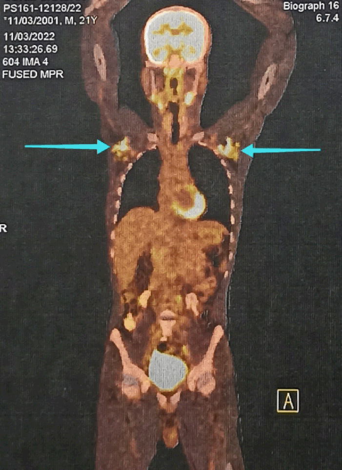
Positron Emission Tomography with Computed Tomography Showing Metabolically Active Axillary Lymph Nodes (Blue Arrow). It was Done After Malignancy was Suspected to Determine the Extent of Spread.
Highlights:Arnab Kundu1, Ramanuj Mukherjee2, Ayan Parichha3, Gouri Mukhopadhyay4
doi: http://dx.doi.org/10.5195/ijms.2024.2147
Volume 12, Number 2: 208-211
Received 05 07 2023; Rev-request 14 09 2023; Rev-request 20 11 2023; Rev-request 23 02 2024; Rev-recd 25 09 2023; Rev-recd 26 11 2023; Rev-recd 29 04 2024; Accepted 06 05 2024
ABSTRACT
Background:Disseminated tuberculosis (TB) is the presence of two or more noncontiguous sites resulting from hematogenous dissemination of Mycobacterium tuberculosis. We report a case of disseminated TB with testicular involvement.
Case:A 21-year-old male patient presented to the outpatient department with bilateral testicular enlargement and tenderness for last six months. It was suspected to be a case of epididymo-orchitis and empirical antimicrobial therapy was initiated. However, ultrasonography findings were inconsistent with epididymo-orchitis. Two weeks later the patient again presented with increased nodularity in the right testes. Non-seminomatous germ cell tumor was suspected. However, tumor markers came back normal. Magnetic resonance imaging revealed enlarged lymph nodes in the right inguinal and retroperitoneal region raising a suspicion of testicular lymphoma. Positron emission tomography with computed tomography showed multiple lymphadenopathies. Histopathology of the left axillary lymph node finally confirmed the diagnosis to be tuberculosis. No drug resistance were found and the patient responded well to anti-tubercular drugs.
Conclusion:Diagnosing disseminated TB is difficult as it mimics conditions, such as infarction, cancer, torsion, etc. Attention to small details is necessary. We faced a similar situation in our patient. The patient went through a myriad of tests before finally being diagnosed with TB. Histopathological study was able to get it whereas cytology could not. Similar and totally opposite cases were found in the literature. This highlights the difficulty and importance of these type of cases.
Disseminated tuberculosis (TB) is common in low and middle-income countries and especially among children below 15 years of age. However, its prevalence in high-income countries is rising due to various risk factors, such as Human immunodeficiency virus (HIV), immunosuppressive medications, organ transplantation, alcohol consumption, and other comorbidities.1
Disseminated TB in an immunocompetent adult comprises less than 2% of all TB cases and up to 20% of all extra-pulmonary TB cases.1 The true global incidence is likely underestimated due to diagnostic challenges.1 It can involve many organs like lymph nodes, liver, bone marrow, kidney, testis, etc.
Clinical presentation varies widely in disseminated TB, ranging from subacute or chronic constitutional symptoms like fever, weight loss, and night sweats to multi-organ failure in severe cases.1 Uncommonly it can present as anorexia and pyrexia of unknown origin.1 Symptoms in children are quite vague, making the diagnosis quite difficult.1 It often mimics various conditions like torsion and infarction (painful testicular mass) or cancer (widespread involvement).
The key to differentiate between various differential diagnoses is by diagnostic tests like imaging, microbiological tests, tissue biopsy, etc. Mantoux test (tuberculin skin test, TST) is often negative in disseminated TB thus not reliable.1 Chest radiograph is often the initial imaging modality, showing characteristic miliary patterns in a majority of cases.1 Other imaging modalities, including high-resolution computed tomography (CT), abdominal ultrasonography, magnetic resonance imaging (MRI), and positron emission tomographic CT (PET-CT), can assist in identifying affected organs and guiding the collection of appropriate specimens for diagnosis.1
Microscopically there are tuberculous granulomas with or without central caseation. Acid-fast bacilli may be found in epithelioid cells or inside caseation. The nonspecific nature of the symptoms poses a challenge in diagnosing the disease.1
Here, we describe a rare case of disseminated TB with testicular involvement, mimicking cancer. This case report highlights the critical need for considering tuberculosis as a diagnosis in testicular masses, emphasizing the diagnostic challenges and atypical presentations mirroring other condition(s), and is crucial for timely and accurate management.
A 21-year-old male presented to the outpatient department (OPD) with complaints of bilateral testicular enlargement for last six months. On examination, both testes were tender, hard and enlarged with loss of testicular sensation as reported by the patient. He was of average build and had no other complaints. He was not currently on any medications and denied any addictions or sexual contact in his life. He denied any history of pulmonary disease or problem. A provisional diagnosis of bilateral epididymo-orchitis was made and the patient was started on empirical antimicrobial therapy. To confirm the diagnosis, urine was sent for routine examination, microscopic examination, and culture which came out normal. Ultrasonography of the scrotum and testis was also advised.
A follow-up visit one week after initiating empiric therapy showed improvement, with reduced pain and tenderness. Two weeks later, the patient again presented to the OPD with a new onset nodularity of the right testis with thickening of the spermatic cord which was not present in the previous OPD visit. Based on scrotal edema, clinical findings suggested a suspected non-seminomatous germ cell tumor, clinically categorized as T4 (As per TNM staging system), with invasion of the spermatic cord and pelvic lymphadenopathy.
However, testicular tumor markers were found to be within the normal limits [LDH - 171U/L (120–246U/L), β-hCG - <0.100mIU/ml (<2.6mIU/ml) and α-fetoprotein - <1.3ng/ml (<8.1ng/ml)]. Later on, MRI of the pelvis and scrotum was done which showed ill-defined T2 hypointensities in both testes along with multiple perifocal satellite hypo-intense lesions. Similar involvement is seen in the epididymis and bilateral spermatic cord (more on the right side). A mild right-sided hydrocele was noted. Also, mildly increased fluid in the left testis was observed. Visible parts of the right ureter were dilated. Enlarged lymph nodes in the right inguinal and retroperitoneal region (mainly the left para-aortic region) were noted. A testicular lymphoma was then suspected based on extensive lymphadenopathy and the absence of any history of tuberculosis.
18FDG whole-body PET-CT was done to determine spread. It showed metabolically active multiple lymphadenopathies (bilateral cervical, axillary, retroperitoneal, iliac and inguinal lymph nodes) and active lymphomatous nodules in bilateral adrenal glands, bilateral epididymis and right testis Figure 1. Then a CT-guided fine needle aspiration cytology (FNAC) of the right inguinal lymph node was done which showed lymphoid cells with foreign body giant cells. No epithelioid tumor or tumor giant cell was seen.
Figure 1.Positron Emission Tomography with Computed Tomography Showing Metabolically Active Axillary Lymph Nodes (Blue Arrow). It was Done After Malignancy was Suspected to Determine the Extent of Spread.
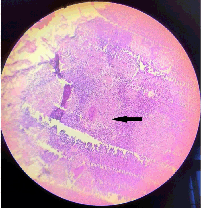
A histopathological examination was advised for confirmation. A left axillary lymph node biopsy was performed and sent for histopathological examination. Preoperative investigations were normal. Mantoux test and triple serology was negative (HIV, Hepatitis B and C). Histopathological examination of axillary lymph nodes showed multiple epithelioid granulomas with Langhans giant cells and foci of caseous necrosis. Figure 2 Histopathological features were in favor of tuberculous lymphadenitis. A diagnosis of disseminated extra-pulmonary TB with testicular involvement was made.
Figure 2.Histopathological Examination of Axillary Lymph Node Showing Caseating Granuloma (black arrow). This Confirmed the Diagnosis of Tuberculosis.
No drug resistance was detected in line probe assay. The patient was started on anti-tubercular treatment of isoniazid, rifampicin, pyrazinamide and ethambutol for two months followed by 4 months of isoniazid, rifampicin and ethambutol. During treatment, the patient was followed up at 1 month, 2 months, 4 months, and 6 months. The patient underwent full remission. Testicular enlargement and nodularity went down and no pain or tenderness were further reported. Lymph node swelling was absent on subsequent visits. The patient now follows up regularly every 6 months Table 1.
Table 1.Histopathological Examination of Axillary Lymph Node Showing Caseating Granuloma (black arrow). This confirmed the diagnosis of tuberculosis.
| Timeframe | Diagnostic tests | Findings | Inferences |
|---|---|---|---|
| Onset | Clinical examination (Initial) | Both testes were tender, hard and enlarged with loss of sensation | Epididymo-orchitis suspected |
| 2 weeks | Clinical examination (Follow up) | Nodularity of right testis and thickening of spermatic cord | Non seminoma germ cell tumor with spermatic cord invasion suspected |
| 1 month | Testicular tumor markers | Normal | Diagnosis of cancer was in doubt |
| 2 months | MRI of pelvis and scrotum | Ill-defined T2 hypointensities in both testes along with multiple perifocal satellite hypo-intense lesions. Similar involvement in the epididymis and bilateral spermatic cord (more on the right side). A mild right-sided hydrocele was noted. Also, mildly increased fluid in the left testis was observed. | Testicular lymphoma was suspected |
| 4 months | 18FDG whole-body PET-CT | Metabolically active multiple lymphadenopathies (bilateral cervical, axillary, retroperitoneal, iliac and inguinal lymph nodes) and active lymphomatous nodules in bilateral adrenal glands, bilateral epididymis and right testis. | Metastasis of suspected testicular lymphoma |
| 7 months | CT-guided Fine Needle Aspiration Cytology of right inguinal lymph node | Lymphoid cells with foreign body giant cells. No epithelioid tumor or tumor giant cell was seen. | Histopathological analysis suggested |
| 9 months | Histopathology of axillary lymph nodes | Multiple epithelioid granulomas with Langhans giant cells and foci of caseous necrosis | Disseminated TB with testicular involvement |
TB can involve many organs like lymph nodes, liver, bone marrow, kidney, testis etc. Acid-fast bacilli may be found in epithelioid cells or inside caseation. Disseminated TB presents with subacute or chronic constitutional symptoms such as fever, weight loss and night sweats. Differential diagnoses include testicular torsion and infarction or cancer. All of these conditions present with testicular enlargement, lump, with or without color changes. Pain is also a common feature but is usually not present in cancer. Also, pain in testicular TB is chronic compared to sudden onset pain torsion and infarction. Specific features of torsion are elevated testis and the absence of cremasteric reflex. Thus, history taking, and clinical examination is important to distinguish between these conditions especially in acute presentations. In testicular masses FNAC is not done due to risk of tumor cell seeding. Only way to do a cytological or histological is an excisional biopsy – This is a significant challenge for the diagnosis of these cases.2
The key to differentiate between various differential diagnoses is by diagnostic tests like imaging, microbiological tests, tissue biopsy, etc. Mantoux test is often negative in disseminated TB thus not so reliable.1 Diagnosis of disseminated TB can be confirmed if any one of the following is present - isolation of tubercle bacilli, positive PCR or Histologic demonstration of caseating granuloma in the biopsy specimen.1 In our case, the symptoms were first interpreted as an inflammatory condition (epididymo-orchitis) which initially improved with later relapse and testicular nodularity. Testicular neoplasms were suspected but it did not fit with the diagnostic tests done later. Ultimately it was diagnosed as a disseminated TB with testicular involvement after histologic evidence of caseating granuloma in the axillary lymph nodes. A case similar to ours was reported in a patient by Namburete EI et al.3 The patient was HIV positive in which case dissemination of TB is common.1,3 However, our patient was immunocompetent without any history of pulmonary disease which makes it unique. Najdawi et al. reported a totally opposite situation in a case of non-seminoma germ cell tumor presenting with dyspnea and cavitary lung lesions which mimicked TB.4 Xiao described a case of testicular TB which was diagnosed after orchiectomy as malignancy was suspected and extensive damage to the testis.5 Mohamed Ali et al. described two cases of testicular tuberculosis from Somalia in immunocompetent patients.6 One had a history of respiratory TB while other one did not. Muttarak M et al. described a case were the diagnosis was based on ultrasonography, no response to conventional antibiotics, and response to first line anti-TB drugs.7 All of this exemplifies the complexity of diagnosing this condition and the differentials.3 In clinical practice, the presentation and history can vary widely as discussed above. Absence of history of any respiratory illness does not necessarily rule out the existence of TB as in our case and the one reported by Xiao, Mohamed Ali et al., and Muttarak M et al. This can be a challenge when a patient presents acutely with the complaints of pain in testes, enlargement and color changes. Histopathology is likely the most effective method to differentiate these conditions as seen in our case and other reported cases.2–5 Medical management by anti-TB drugs is the mainstay of treatment but sometimes surgical management becomes necessary.8 This case contributes to the existing literature on disseminated TB and will help clinicians to be more aware and cautious while diagnosing conditions with testicular lesions.
A 21-year-old male presented with bilateral testicular enlargement. Initially it was diagnosed as epididymo-orchitis. Further investigations revealed nodules, thickening and lymphadenopathy. Later on MRI was done and testicular lymphoma was suspected. The PET-CT confirmed multiple active lymphadenopathies and nodules. Axillary lymph node biopsy was done which lead to a diagnosis of disseminated extra-pulmonary tuberculosis with testicular involvement. The patient responded well to anti-tubercular treatment. This case highlights the complexity of diagnosing disseminated TB and underscores the importance of histopathological examination for confirmation in atypical presentations. Similar and opposite cases have been reported in the literature exemplifying the complexity and importance of these types of cases. Recognizing these cases is essential as it reduces the risk of complications if treatment is initiated early.
None
The Authors have no funding, financial relationships or conflicts of interest to disclose.
Conceptualization: AK, RM. Data Curation: AK. Methodology: AK, AP. Project Administration: RM, GM. Resources: AK, RM, AP. Supervision: RM, GM. Validation: RM, GM. Writing - Original Draft: AK, AP. Writing - Review Editing: AK, RM, AP, GM
1. Khan FY. Review of literature on disseminated tuberculosis with emphasis on the focused diagnostic workup. J Family Community Med. 2019;26(2):83–91.
2. Bhargava A, Davenport C, Gibbons N, McConkey S. TB or not tb?: A case of isolated testicular TB with scrotal involvement. Ir J Med Sci. 2008;178(2):231–3.
3. Namburete EI, Di Gennaro F, Jose Maria C, Fiore Bavaro D, Brindicci G, Lattanzio R, et al. Uncommon testicular localization of Disseminated TB: a case report from Mozambique. New Microbiol. 2019;42(3):184–187.
4. Najdawi F, Means M, Didde R, Feloney M. Testicular cancer presenting as disseminated tuberculosis: A case report. Ann Med Surg (Lond). 2021;72:102975.
5. Xiao K. Testicular Tuberculosis. N Engl J Med. 2023;389(8):e13.
6. Mohamed Alı A, Doğan A, Ali MA, Çakmak BS. Testicular Tuberculosis: Two Rare Case Report. Int Med Case Rep J. 2023;16:339–343.
7. Muttarak M, Peh WC. Case 91: Tuberculous epididymo-orchitis. Radiology. 2006;238(2):748–51.
8. Carl P, Stark L. Indications for surgical management of genitourinary tuberculosis. World J Surg. 1997;21(5):505–10.
Arnab Kundu, 1 Third-year MBBS student, R.G. Kar Medical College and Hospital, Kolkata, India.
Ramanuj Mukherjee, 2 MBBS, MS, DNB, MNAMS, MRCS, FMAS, FACS. Professor, Department of General Surgery, R.G. Kar Medical College and Hospital, Kolkata, India.
Ayan Parichha, 3 MBBS. Intern, R.G. Kar Medical College and Hospital, Kolkata, India.
Gouri Mukhopadhyay, 4 MBBS, MS. Adyar Cancer Institute, Chennai, India.
Correspondence: Arnab Kundu. Address: Medical College Kolkata, 88, College St, College Square, Kolkata, West Bengal 700073. Email: karnab48@gmail.com
Editor: Francisco J. Bonilla-Escobar; Student Editors: Michael V. Tavolieri & Carlos de la Cruz-de la Cruz; Proofreader: Amy Phelan; Layout Editor: Julián A. Zapata-Ríos; Process: Peer-reviewed
Cite as Kundu A, Mukherjee R, Parichha A, Mukhopadhyay G. Disseminated Tuberculosis with Testes Involvement: An Intriguing Case Report. Int J Med Stud. 2024 Apr-Jun;12(2):208-211.
Copyright © 2024 Arnab Kundu, Ramanuj Mukherjee, Ayan Parichha, Gouri Mukhopadhyay
This work is licensed under a Creative Commons Attribution 4.0 International License.
International Journal of Medical Students, VOLUME 12, NUMBER 2, May 2024