Contrast-Enhanced T2-Weighted MRI Scan; (A) Coronal, (B) Axial, and (C) Sagittal Sections of Left Hand.

Highlights:Made Bramantya Karna1, Richard Christian Suteja2
doi: http://dx.doi.org/ijms.2024.2486
Volume 12, Number 3: 338-344
Received 17 12 2023; Rev-request 23 02 2024; Rev-recd 28 02 2024; Accepted 14 07 2024
ABSTRACT
Background:Glomus tumors are rare benign hamartomas of the glomus body that occur mostly – though not limited to - the distal phalanxes of the digits. This article provides a real-life example of successful rare disease identification and treatment. It also provides a guideline that may help serve as future guidance on diagnosis and treatment.
Case:A 41-year-old male came to our hospital presenting with a chief complaint of episodes of throbbing pain, which occurred spontaneously on the left thumb for the past two years. The pain had worsened in the past two weeks. The patient was positive for Hildreth's, Love's pin, and cold sensitivity tests. The previous x-ray showed no abnormalities in the left thumb. MRI found a hyperdense subungual lesion in the dorsal interphalangeal joint of the left thumb. We then performed an excision using the transungual approach. Histopathological findings found a relatively well-circumscribed lesion of the glomus apparatus absent of abnormal mitosis and necrosis. Two months after the excision, the patient reported no symptoms of recurrency, nail deformity, or other adverse outcomes.
Conclusion:Patients typically present a chief complaint of chronic paroxysmal throbbing nail pain that persists for years, increases following exposure to cold environments, and is disproportionately exacerbated with the slightest touch. Hildreth's, Love's Pin, and cold sensitivity tests are special examinations that elicit or suppress pain. As with most benign tumors, complete excision usually yields good results. Adequate knowledge about diagnostic methods will help patients achieve early intervention and cure.
Glomus tumors are rare benign hamartomas of the glomus body that occur mostly – though not limited to - the distal phalanxes of the digits.1,2 Patients often present to the clinic with symptoms of paroxysmal pain in tissues below the nail.3-5 The severity of the pain is proportional to the size of the tumor. The pain is exacerbated when touched or exposed to cold environments.3-5 Studies report that glomus tumors only form about 1–5% of all hand tumors.2 In most patients, initial pain is generally tolerable due to its paroxysmal and non-severe nature, However, as the tumor grows, the pain may be felt more frequently, with each episode being progressively painful. Furthermore, patients with chronic pain may develop a higher threshold when tolerating pain. This creates a delay from the patient's side in seeking treatment.
Studies report a mean length of time from initial symptom onset to diagnosis of around 7 years.6 Aside from delays from the patient's side, it is reasonable to suspect our current knowledge on the disease. The lack of understanding and experience due to less to no contact to this disease during physician's training period may well contribute to the number of misdiagnoses. Albeit its lack of emergency, glomus tumors may reduce patient's quality of life, as there may be some limitation when performing specific movements such as gripping.
A knowledgeable and keen physician should be able to suspect a glomus tumor based on presented clinical symptoms. This is due to the disease's generally unique and distinguishable symptoms. Additionally, several specific tests may be performed to confirm the diagnosis. A transillumination test and MRI scan may also be performed on the affected digit.7,8 However, a negative transillumination and MRI scan result do not necessarily exclude the possibility of glomus tumors when specific symptoms are present.7,9-11 Prompt treatment may restore lost quality of life by allowing the patient to function as normal. In this article, we provide a case of successful glomus tumor diagnosis and removal. This case is uncommon, and therefore may serve as a learning material for medical students. This article is written while adhering to CARE guidelines.12 This article will also propose a diagnostic scheme to help physicians diagnose glomus tumors.
A male in his 40s presented with a chief complaint of pain under the nail of his left thumb for the past two years prior to coming to our hospital, which worsened in the past two weeks. The pain was isolated and only felt on his left thumb. The patient described the pain as episodes of throbbing pain that occurred spontaneously. The patient felt that pain episodes were more prevalent during certain times of the year, and the throbbing felt greater when exposed to cold air from the refrigerator.
The patient reported that he had previously sought care from multiple doctors over the two years but to no avail. There are neither accompanying symptoms nor significant past, family, or social histories. There is also no history of trauma in the said location. Upon inspection, we found no discoloration, swelling, or deformity. However, tenderness was present and was localized to the dorsal side of the distal interphalangeal joint. Albeit localized within the joint, passive and active movement was not restricted. The presentation of paroxysmal, throbbing pain, exacerbated following exposure to a cold environment, warrants further provocation and transillumination tests, which, in our case, showed positive results (Table 1).
Table 1.Key Symptoms and Diagnostic Findings.
| Key Symptoms |
|
| Key Diagnostic Findings |
|
The patient came to our clinic with left thumb x-ray results from a scan he underwent after an appointment with his previous doctor. The x-ray showed no abnormalities in the left thumb and, in general, his left hand. A complete blood count found no significant results. Contrast-enhanced T2-weighted MRI scan found a well-demarcated 1.7 cm x 0.6 cm x 0.6 cm hyperdense lesion in the dorsal interphalangeal joint of the left thumb separate from the bone Figure 1.
Figure 1.Contrast-Enhanced T2-Weighted MRI Scan; (A) Coronal, (B) Axial, and (C) Sagittal Sections of Left Hand.

Initial findings and epidemiologic study made us suspect an early-formed ganglion cyst because it is one of the most prevalent soft tissue masses in hand surgery.13 However, the feeling of pain was not typical of ganglion cysts and warrants further investigation. This makes our working diagnosis the glomus tumor of the hand, and further pathological examination was required to confirm this diagnosis.
We performed a surgical excision of the benign lesion using the transungual approach. This approach was chosen due to the location of the tumor as shown by MRI. Under local digital block, a finger tourniquet was used to exsanguinate the finger subjected to surgery. Antiseptic preparation was done on the surgical site, and then an incision was made along the lateral aspect of the proximal nail fold, followed by nail plate extraction. An exposed nail bed was then incised longitudinally right above the lesion site, allowing tumor exposure. Following the excision of the encapsulated tumor, the nail bed was then sutured, and the lifted nail was replaced on top of the nail bed. The nail was then sutured with the proximal nail fold, and wound dressing with gauze and antibiotic ointment was changed daily until the wound was dry, after which it was kept open.
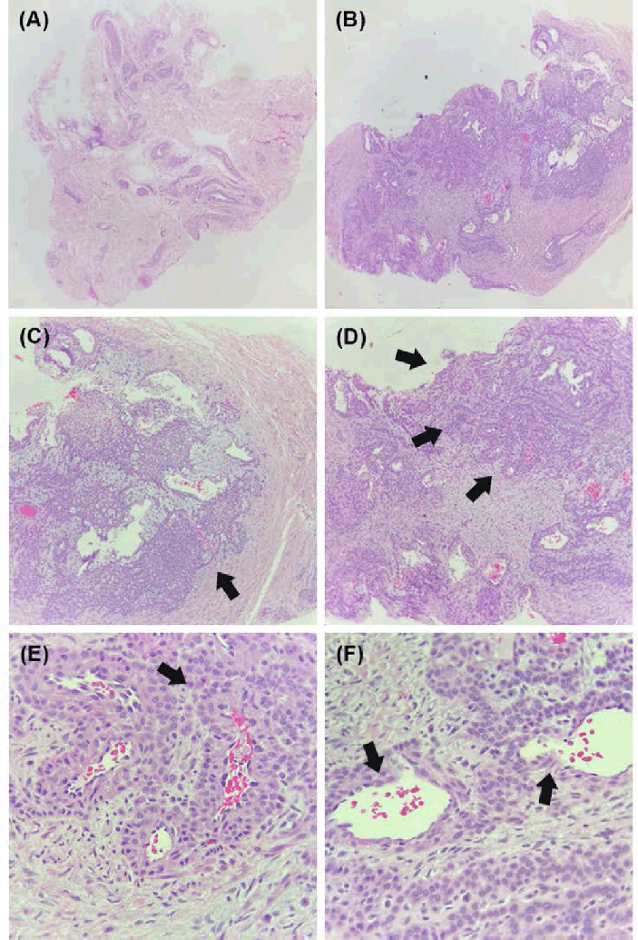
Following surgery, we successfully extracted two greyish-white tissues sized 0.4 cm x 0.2 cm x 0.2 cm and 0.3 cm x 0.2 cm x 0.2 cm, respectively. These tissues were then immersed in a 10% NBF (neutral buffered formalin). The excised tissue's pathological findings Figure 2 correspond to the conventional morphology of the type II glomus tumor.
Figure 2.Histopathological Findings of Lesions.

Two months following the excision, we monitored for any possibility of recurrence or post-treatment adverse effects. The patient reported no recurrence of symptoms such as pain, tenderness, or local inflammation. No nail deformity or any other adverse outcomes were reported. The patient was then able return to his work without any meaningful limitations. He can now carry things that require extensive use of his thumb symmetrically with his left and right hand. Provocation tests were performed again, this time showing negative results.
In our study, the patient presented symptoms which correlated to two of the three tests specific for the diagnosis of glomus tumor: paroxysmal subungual throbbing pain exacerbated when exposed to cold environments. These symptoms present in 90–100% of patients, as mentioned in several other studies which report 10 or more patients with digital glomus tumor.14,15 Patients may also present with other symptoms such as nail disfigurement or discoloration.14,15 However, these were not present in our patient. Following confirmation through physical and supporting examination, we decided that excision via transungual approach was the best surgical method due to the tumor being more centrally located. Albeit being the method which offers the best visualization and exposure, this technique is the most disruptive to the nail bed, increasing chances of complications such as inaesthetic nail, nail ridging, and nail splitting.14,16,17 Other options include the lateral and volar approach, which were more restricted in visualization but are less disruptive towards the nail bed.14,18,19 Structurally, a glomus apparatus consists of a single afferent arteriole and efferent venule, connected through multiple arteriovenous anastomoses (Suquet-Hoyer canals), altogether encapsulated with α-actin containing muscle fibers and glomus bodies.20 The afferent arterioles are formed by endothelium, internal elastic lamina, and pre-glomic muscle cells, while efferent venules comprise a thin endothelium layer.20 The glomus apparatus physiologically functions as a contractile neuro-myo-arterial unit that autonomically thermoregulates body temperature through shunting and maximizing blood flow within the cutaneous microvasculature.20 On exposure to cold temperatures, a marked increase in norepinephrine results in constriction of blood vessels, shunting blood flow towards the apparatus.21 Decrease of peripheral blood flow causes a reduction in convection-mediated heat loss. Hyperplasia of either one of the components within the glomus apparatus (i.e., blood vessel, glomus body, muscle) creates a condition known as a glomus tumor.22 Generally, there are three types of glomus tumors based on their histopathological differences.
Although strictly differentiated into distinct types, there are cases where patients may present with multiple histopathological types concurrently.23,24 The three main classifications are: (1) hyaline-mucoid type, (2) solid type, and (3) angiomatous type.23,24 Hyaline-mucoid type glomus tumor is characterized by increased hyalinized connective tissue interspersed within islands of glomus cells and vascular spaces.23,24 Solid-type glomus tumor is characterized by glomus cell masses and reduced vascular and muscle space.23,24 This type was previously called the ‘typical’ glomus tumor histology. An increase in intracapsular vasculature characterizes the angiomatous glomus tumor and is the rarest among the three types.23,24
Although glomus apparatuses can be found in various parts of the body, a typical glomus tumor occurs as a solitary, benign lesion typically located on subungual tissues of the digits.1 Patients typically present a chief complaint of chronic paroxysmal throbbing nail pain, which persists for years, as it is often ignored or, sometimes, misdiagnosed.3-5 This pain increases following exposure to cold environments and is disproportionately exacerbated due to the slightest touch.3-5 In some cases, the tumor may present together with a bluish-pink nodule or longitudinal nail splitting.4,5
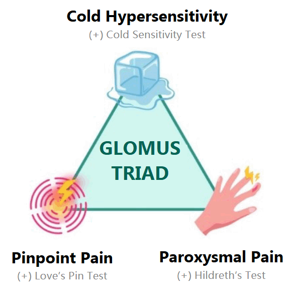
Owing to the relatively unique symptoms, the differential diagnosis of glomus tumor must be added following the presentation of paroxysmal throbbing pain, disproportionate pinpoint pain, and cold hypersensitivity, as shown in Figure 3. Physicians generally perform three special examinations that aim to elicit or suppress pain following exposure to certain conditions: Hildreth's Test, Love's Pin Test, and cold sensitivity Test. Love's pin test is performed by applying pressure to the mass using a rounded-tip object such as a ballpoint pen, pinhead, or the end of a paperclip.25 Love's pin test is positive if the pain is elicited after applying pressure, a refractory hand withdrawal is demonstrated.25,26 Love's pin test is also commonly used to roughly localize the lesion by identifying areas exhibiting the most severe pain.26 Hildreth's test is performed by exsanguinating the affected digit using a tourniquet applied at the base of the digit.27 The physician will then observe any signs of reduction in pain and tenderness, indicating a positive Hildreth test.27 A surge of sudden pain may occur following the release of the tourniquet. Giele (2002) observed that this test was 92% sensitive and 91% specific to glomus tumors.27 Reduced or absence of pain by repeating Love's pin test following application of tourniquet (sharpened Hildreth's test) also indicates a positive result.26 Cold sensitivity test exposes the affected digit to cold environments, usually by applying an ice pack or cubes directly above the suspected lesion site. Induction of pain indicates a positive cold sensitivity test.28 These tests can be seen in Figure 4.
Figure 3.Glomus Triad.

Provocation Tests: (A) Love's Pin Test, (B) Hildreth's Test, (C) Cold Sensitivity Test.

Some studies also recommended the use of a transillumination test.7 The test is performed by placing a flashlight on the palmar side of the affected digit and then looking for any inconsistencies in opacity within the nail plate and bed.7 This method is an easy, non-invasive, cost-effective adjuvant diagnostic tool.7 In addition to specific physical examinations, glomus tumors can be diagnosed using high-resolution magnetic resonance imaging (MRI). In a contrast-enhanced MRI scan, a typical glomus tumor is a hyperintense lesion with a well-demarcated border.8 However, it must be noted that a negative MRI result on a clinically suspected glomus tumor patient does not exclude the possibility of a hidden tumor within the digits.9-11
Differential diagnoses include ganglion cysts, subungual melanoma, and subungual squamous cell carcinoma. However, ganglion cysts should be characterized as a non-painful lump except if they compress a nearby nerve.29 In our case, the lesion was found to be more centrally-located and was not situated in a location highly possible for nerve compression.30 Patients with ganglion cysts may also report preceding traumatic event or repetitive stretching of nearby capsular and ligamentous structure which stimulates production of tissue hyaluronic acid.31,32 T2-weighted MRI of ganglion cysts may show hyperdense lesion(s) similar to glomus tumor. However, lesions are usually situated above joints.30 Subungual melanoma was also a possible differential diagnosis. However, it tends to manifest with nail discoloration, which was not present in our patient.33 As they are more malignant, lesions which had a longer onset, such as in our patient, should include bone involvement, nail onycholysis, and erosion/ulceration of the nail bed.34,35 This was not observed in our patient's X-ray and MRI results. Similar to subungual squamous cell carcinoma, lesions with a longer onset usually present larger mass with bony involvement, nail onycholysis, nail dystrophy, and other more destructive symptoms which were not present in our patient.36 Keratotic/verrucous lesion may also be present on the nail bed following nail plate loss.36
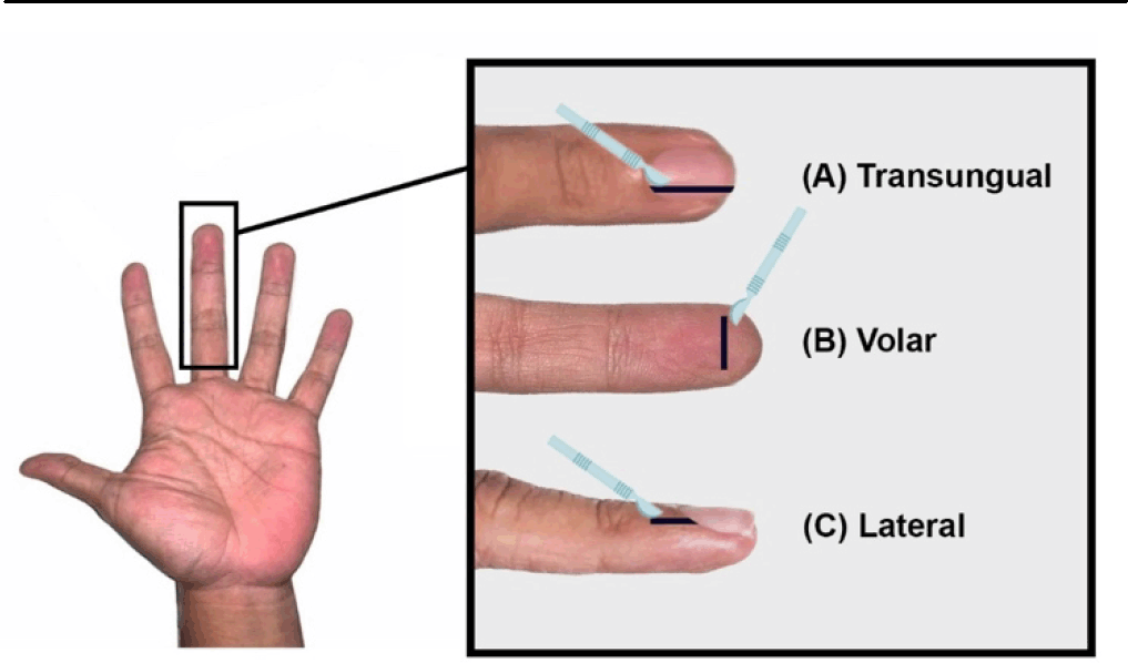
As with most benign tumors, complete excision usually yields good results. There are several approaches to excision: the transungual, lateral, and volar approaches (shown in Figure 5.1,17-19,37 A transungual approach generally involves an incision along the lateral aspect of the proximal nail fold to allow nail plate extraction. Another longitudinal incision then allows tumor excision. The nail bed is then sutured again, and the lifted nail is be replaced on top of the nail bed. A lateral approach involves. making an incision along the side of the nail or finger, which allows for excision of the tumor with less disruption to the nail bed, potentially reducing the risk of nail deformity. The transungual approach generally provides better exposure to the tumor, increasing certainty towards complete excision with the expense of nail deformity due to massive manipulations.17 Transungual approach is generally used for the typical centrally-situated subungual glomus tumor. A lateral approach involves a lateral high incision to the side nearer the tumor. The incision was then extended distally to cover the pulp.1
Figure 5.Various Approaches in Surgical Excision of Digital Glomus Tumor (A) Transungual Approach, (B) Volar Approach, (C) Lateral Subperiosteal Approach.

Thereafter, a deep dissection was performed, exploring subperiosteally to the distal phalanx, hence raising a dorsal flap of the nail matrix, nail bed, and nail plate in one single unit, and keeping the nail bed-to-plate integrity.1 The magnitude of elevation depend on the proximity of the tumor.1,37 A modification to this technique is the lateral subperiosteal approach, which omitted further incision towards the pulp.37 This approach is advantageous as it is quicker and protects the nail against post-surgery deformity.37 However, studies report that this approach may sometimes cause incomplete excision and early tumor recurrence. This approach is generally used for a more laterally-situated subungual glomus tumor.17 The volar approach involves an incision on the palmar side of the digits and is used only when the mass is situated more towards the palmar or volar side.18,19
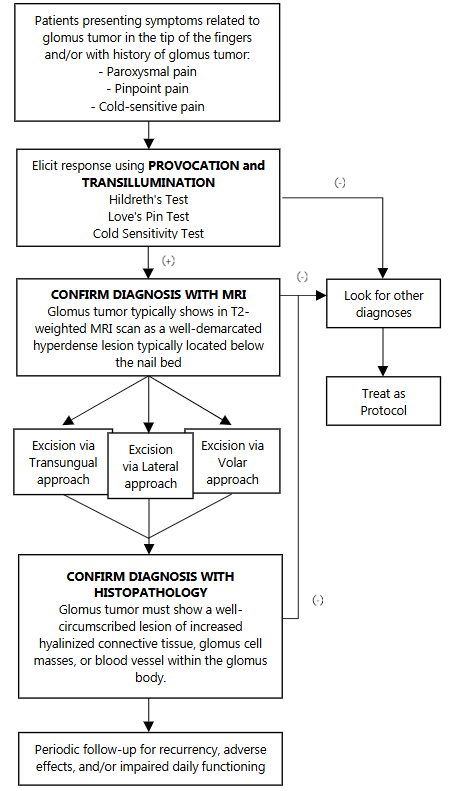
Based on the literature review and our personal experience in this case, we propose a diagnostic and treatment workflow, as shown in Figure 6. We suggest performing all provocation tests alongside transillumination test even when only one symptom of the triad is present, especially on patients with a history of glomus tumor. The provocation procedures are relatively easy and cheap, sparing the patient of further loss.
Figure 6.Diagnostic and Therapeutical Algorithm for Glomus Tumors.

Although the typical glomus tumor often occurs as a benign solitary nodule in the digits, it is anatomically possible for a glomus tumor to occur in other parts of the body, as other parts contain glomus bodies, too. These glomus tumors outside of the digits usually present with malignant-like characteristics such as multiple and large nodules, as well as atypical mitotic figures, infiltrative, and can metastasize.38,39 These types of glomus tumors have a higher recurrence rate following excision.39 Although this study reports a successful rare tumor removal, data regarding long-term (>12 months) recurrency or adverse effects follow up was not available. Literature reported that recurrence occurs in up to 18% of cases.40 However, only tumors recurring after 24 months post-excision can be regarded as true recurrence rather than pseudo-recurrence which occurs due to inadequate excision.41
In conclusion, glomus tumors are rare benign hamartomas of the glomus body that occur mostly - though not limited to - the distal phalanxes of the digits. Patients typically present with a chief complaint of chronic paroxysmal throbbing nail pain that persists for years, increases following exposure to cold environments, and is disproportionately exacerbated due to the slightest touch. Hildreth's, Love's Pin, and cold sensitivity tests were special examinations that elicit or suppress pain. We suggest performing all provocation tests and the transillumination test even when only one symptom of the triad is present, especially on patients with a history of glomus tumor. The provocation procedures are relatively easy and cheap, sparing the patient of further loss. As with most benign tumors, complete excision usually yields good results. Adequate knowledge about diagnostic methods will help patients achieve early intervention and resolvent.
Upon follow up, the patient stated an almost 100% functional daily activity recovery. The patient reported that he is satisfied with the results, both cosmetically and functionally. Provocation tests specific for glomus tumor were redone and found to be negative.
Glomus tumors are rare benign hamartomas of the glomus body that occur mostly – though not limited to - the distal phalanxes of the digits. Patients typically present a chief complaint of chronic paroxysmal throbbing nail pain that persists for years, increases following exposure to cold environments, and is disproportionately exacerbated due to the slightest touch. Hildreth's, Love's Pin, and cold sensitivity tests are special examinations that elicit or suppress pain. We suggest performing all provocation tests and transillumination test even when only one symptom of the triad is present, especially on patients with a history of glomus tumor. The provocation procedures are relatively easy and cheap, sparing the patient of further loss. As with most benign tumors, complete excision usually yields good results. Adequate knowledge about diagnostic methods will help patients achieve early intervention and resolvent.
None
The Authors have no funding, financial relationships or conflicts of interest to disclose.
Conceptualization: MBK. Data Curation: MBK. Investigation: MBK, RCS. Methodology: MBK, RCS. Project Administration: MBK, RCS. Resources: MBK, RCS. Supervision: MBK. Validation: MBK. Writing – Original Draft: MBK, RCS. Writing – Review Editing: MBK.
1. Morey VM, Garg B, Kotwal PP. Glomus tumors of the hand: review of literature. J Clin Orthop Trauma. 2016;7(4):286–91.
2. Jawalkar H, Maryada VR, Brahmajoshyula V, Kotha GKV. Subungual glomus tumors of the hand treated by transungual excision. Indian J Orthop. 2015;49(4):403–7.
3. Bordianu A, Zamfirescu D. The hidden cause of chronic finger pain: glomus tumor—a case report. J Med Life. 2019;12(1):30–3.
4. Newman MJ, Pocock G, Allan P. Glomus tumour: a rare differential for subungual lesions. BMJ Case Rep. 2015;2015.
5. Carroll RE, Berman AT. Glomus tumors of the hand: review of the literature and report on twenty-eight cases. J Bone Joint Surg Am. 1972;54(4):691–703.
6. Kim DH. Glomus tumor of the fingertip and MRI appearance. Iowa Orthop J. 1999;19:136–8.
7. Tsiogka A, Belyayeva H, Sianos S, Rigopoulos D. Transillumination: a diagnostic tool to assess subungual glomus tumors. Skin Appendage Disord. 2021;7(3):231–3.
8. Al-Qattan MM, Al-Namla A, Al-Thunayan A, Al-Subhi F, El-Shayeb AF. Magnetic resonance imaging in the diagnosis of glomus tumors of the hand. J Hand Surg Am. 2005;30(5):535–40.
9. Bargon CA, Mohamadi A, Talaei-Khoei M, Ring DC, Mudgal CS. Factors associated with requesting magnetic resonance imaging during the management of glomus tumors. Arch Bone Jt Surg. 2019;7(5):422–8.
10. Chou T, Pan SC, Shieh SJ, Lee JW, Chiu HY, Ho CL. Glomus Tumor: Twenty-Year Experience and Literature Review. Ann Plast Surg. 2016;76 Suppl 1: S35–40.
11. Fazwi R, Chandran PA, Ahmad TS. Glomus tumour: a retrospective review of 15 years' experience in a single institution. Malays Orthop J. 2011;5(3):8–12.
12. Equator Network. CARE Checklist English 2013. Available from: https://www.care-statement.org/checklist. Cited Jun 26, 2023.
13. Zampeli F, Terzidis I, Bernard M, Ochi M, Pappas E, Georgoulis A. Ganglion cyst. In: StatPearls [Internet]. Treasure Island (FL): StatPearls Publishing; 2023 Jul 17 [cited Dec 17, 2023]. Available from: https://www.ncbi.nlm.nih.gov/books/NBK470168/
14. Shin DK, Kim MS, Kim SW, Kim SH. A painful glomus tumor on the pulp of the distal phalanx. J Korean Neurosurg Soc. 2010;48(2):185–7.
15. Shepherd JT, Rusch NJ, Vanhoutte PM. Effect of cold on the blood vessel wall. Gen Pharmacol. 1983;14(1):61–4.
16. Masson P. Le glomus neuromyoartérial des régions tactiles et ses tumeurs. Lyon Chir. 1924;21:257–80.
17. Tuncali D, Yilmaz AC, Terzioglu A, Aslan G. Multiple occurrences of different histologic types of the glomus tumor. J Hand Surg Am. 2005;30(1):161–4.
18. Looi KP, Teh M, Pho RW. An unusual case of multiple recurrence of a glomangioma. J Hand Surg Br. 1999;24(3):387–9.
19. Love JG. Glomus tumors: diagnosis and treatment. Proc Staff Meet Mayo Clin. 1944;19:113–6.
20. Tang CYK, Tipoe T, Fung B. Where is the lesion? Glomus tumours of the hand. Arch Plast Surg. 2013;40(5):492–5.
21. Yii NW, Elliot D. Hildreth's test is a reliable clinical sign for the diagnosis of glomus tumors. J Hand Surg Br. 2002;27(2):157–8.
22. Netscher DT, Aburto J, Koepplinger M. Subungual glomus tumor. J Hand Surg Am. 2012;37(4):821–3.
23. Kabakas F, Ugurlar M, Ozcelik I, Mersa B, Purisa H, Sezer I. Surgical treatment of subungual glomus tumors: experience with lateral subperiosteal and transungual approaches. Hand Microsurg. 2016;5(2):70–4.
24. Garg B, Machhindra MV, Tiwari V, Shankar V, Kotwal P. Nail-preserving modified lateral subperiosteal approach for subungual glomus tumour: a novel surgical approach. Musculoskelet Surg. 2016;100(1):43–8.
25. Mortada H, AlRabah R, Kattan AE. Unusual Location of Pulp Glomus Tumor: A Case Study and Literature Review. Plast Reconstr Surg Glob Open. 2022;10(3):e4206.
26. Senhaji G, Gallouj S, El Jouari O, Lamouaffaq A, Rimani M, Mernissi FZ. Rare tumor in unusual location–glomus tumor of the finger pulp (clinical and dermoscopic features): a case report. J Med Case Rep. 2018;12(1):1–5.
27. Binesh F, Akhavan A, Zahir ST, Bovanlu TR. Clinically malignant atypical glomus tumour. BMJ Case Rep. 2013;2013.
28. Folpe AL, Fanburg-Smith JC, Miettinen M, Weiss SW. Atypical and malignant glomus tumors: analysis of 52 cases, with a proposal for the reclassification of glomus tumors. Am J Surg Pathol. 2001;25(1):1–12.
Made Bramantya Karna, 1 MD, PhD. Hand Division, Orthopaedics and Traumatology Department, Faculty of Medicine, Udayana University/Prof. dr. IGNG Ngoerah General Hospital, Denpasar, Bali.
Richard Christian Suteja, 2 Fourth-year medical student, Faculty of Medicine, Udayana University, Denpasar, Bali.
About the Author: Richard Christian Suteja is a fourth-year medical student at the Faculty of Medicine, Udayana University, Denpasar, Bali. He is working to finish his degree at Prof. dr. IGNG Ngoerah General Hospital, Bali, Indonesia.
Correspondence: Richard Christian Suteja. Address: 86G9+HCW, Jl. P.B. Sudirman, Dangin Puri Klod, Kec. Bali 80232, Indonesia. Email: bram.ortho@outlook.com
Editor: Francisco J. Bonilla-Escobar; Student Editors: Viviana Cortiana & Rachna Shekhar; Proofreader: Laeeqa Manji; Layout Editor: Julian A. Zapata-Rios
Cite as Made Bramantya K, Suteja RC. Successful Subungual Glomus Tumor Removal: A Case Report and Future Guidance on Diagnosis and Treatment. Int J Med Stud. 2024 Jul-Sep;12(3):338-344.
Copyright © 2024 Made Bramantya Karna, Richard Christian Suteja
This work is licensed under a Creative Commons Attribution 4.0 International License.
International Journal of Medical Students, VOLUME 12, NUMBER 3, July 2023