Case Report
Complex Regional Pain Syndrome, an Important Differential Diagnosis in Sports Injuries:
a Case Report
Carlos Cabrera-Ubilla1, Germán Cueto2, Christian Lucas3
doi: http://dx.doi.org/ijms.2024.2522
Volume 12, Number 4: 465-467
Received 17 01 2024;
Rev-request 15 07 2024;
Rev-recd 16 07 2024;
Accepted 16 09 2024
ABSTRACT
Background:
Complex regional pain syndrome (CRPS) is a disproportionate and persistent, regional
pain related to a minor trauma. Although CRPS is not an infrequent condition its pathophysiology
remains unknown and leading to underdiagnosis or late diagnosis. The diagnosis is
clinical, according to Budapest criteria of the International Association for the
Study of Pain. Bone scintigram is the most effective test to support the diagnosis.
The aim of this article is to discuss the importance of clinical suspicion for an
early CRPS diagnosis in a sprain's young athlete clinical case.
The Case:
We present the case of a sixteen-year-old male patient with no medical history who
suffered two minor ankle injuries in the right foot. The patient developed severe
and persistent pain associated with vasomotor, sudomotor and trophic abnormalities.
He remained undiagnosed for 10 months until CRPS diagnosis confirmation supported
by a bone scintigram. He received multiple treatments until spontaneous remission
in the fourth year of evolution.
Discussion:
CRPS poses a diagnostic challenge that requires early suspicion to improve treatment
outcomes and prognosis. Maintaining a high index of clinical suspicion is crucial,
and CRPS should be considered in the evaluation of any persistent pain sport-related
injury. Despite extensive research on CRPS conducted in recent decades, this condition
may still be unfamiliar to many healthcare providers. Increasing awareness of CRPS
among medical professionals can facilitate timely and accurate diagnosis, which is
essential for effective management.
Highlights:
- CRPS is a disproportionate and persistent, regional pain related to a minor trauma.
- CRPS represents a diagnostic challenge that should always be considered in the context
of a persistent pain sport-related injury.
- Despite extensive research on CRPS, it may still be unfamiliar to the medical community.
Raising awareness is crucial to aiding healthcare professionals in making timely and
accurate diagnoses.
Introduction
Complex regional pain syndrome (CRPS) is defined as an array of painful conditions
characterized by a disproportionate continuing regional pain.1 The incidence varies widely between 5 to 26 per 100,000 inhabitants.2 It is caused mainly after a local trauma, most frequently a fracture or sprain.3 In the last two decades, CRPS research has been largely conducted. However, the pathophysiology
mechanism is not yet fully understood.4
The diagnosis is clinical by the modified diagnosis criteria of the International
Association for the Study of Pain (IASP) or Budapest criteria, which established the
CRPS as an exclusion diagnosis characterized by a continuing pain disproportionate
to any incited event associated with sensory (hyperalgesia and/or allodynia), vasomotor
(temperature and/or skin color asymmetry), sudomotor (edema and/or sweating) and motor/trophic
changes in a limb region, that doesn't follow any specific nerve territory or dermatome.5
CRPS continues to be a diagnosis challenge, with a significant delay in the diagnosis,
which worsened the prognosis and treatment response.6 Considering that the precipitating event in CRPS is often represented by an injury,
this condition should be relevant for athletes.3 The aim of this article is to discuss the importance of clinical suspicion for an
early CRPS diagnosis in a sprain's young athlete clinical case.
The Case
We present the case of a sixteen-year-old male patient with no medical history, who
suffered two consecutives grade one right ankle sprain while playing soccer. He had
poor response to 15 kinesiotherapy sessions. Afterwards, he progressed with a burning
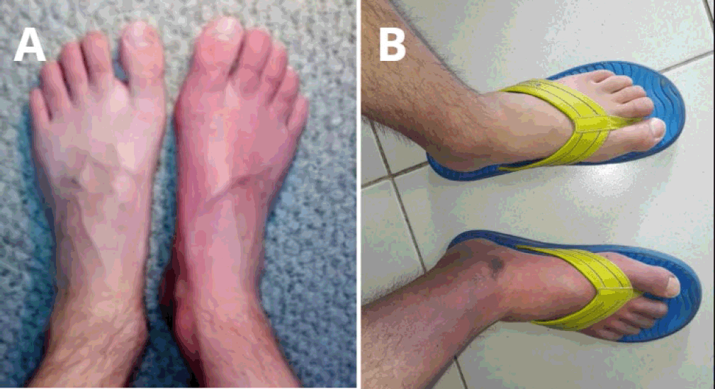
pain, increased volume and red-purple erythema triggered by the heat. Physical examination
Figure 1.A showed mild edema, erythema, allodynia, higher temperature and hair loss in the right
foot, without motor alterations. A right foot ultrasound was performed, revealing
a partial anterior talofibular ligament rupture and a mild peroneal tendonitis. Based
on these findings, the patient continued with kinesiotherapy.
Figure 1
Patient's Physical Examination at Third and Tenth Month of Evolution.

Legend: Physical examination at the third month (Figure 1.A): asymmetrical skin changes, with greater erythema in the right foot. Physical examination
at the tenth month (Figure 1.B): increase of asymmetrical erythema, edema and hair loss in the right foot.
Due to pain persistence at the sixth month, the case study was complemented with vascular
and rheumatological tests, which were negative. Additionally, it was performed a right
ankle MRI, which reported the presence of calcaneal bone edema. Therefore, new kinesiotherapy
sessions were indicated. At the tenth month, the symptoms worsened, increasing the
affected area until middle leg Figure 1.B. Since symptoms could not be explained by the imaging findings, a lower member bone
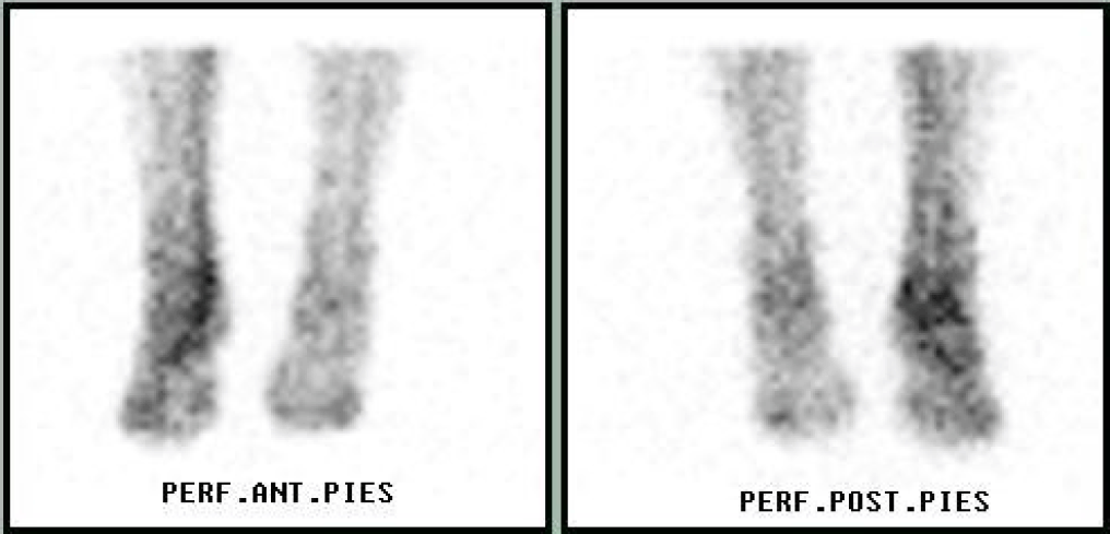
scintigram was requested Figure 2. The exam reported a moderate increase in arterial flow and diffuse hyper uptake
in right foot and ankle, confirming the diagnosis of a CRPS.
Figure 2
Lower Member Bone Scintigram.

Legend: An anterior and posterior vision of a lower member bone scintigram evidence a moderate
increase in arterial flow and diffuse hyperuptake in right foot and ankle. These findings
correlated with the autonomic dysfunction in CRPS, leading to blood flow abnormalities
The patient began treatment with pregabalin and duloxetine in ascending doses until
75 mg every 12 hours and 60 mg per day, respectively. In addition, he received psychotherapy
for pain management. Symptoms worsened after one year of treatment. Further therapeutics
options were considered. The patient underwent 5 sympathetic blocks sessions, not
being able to complete the 10 recommended sessions. Symptoms remained stable and the
patient abandoned medical therapies in the third year. He explored alternative medicines
such as acupuncture, phytotherapy, mindfulness, among others, without significant
relief. Medical follow-up continued and during the fifth year of disease there was
a progressive decrease in symptoms until remission. Only mild vegetative symptoms
persisted but there was an absence of pain and full recovery of functionality.
Discussion
We presented the case of a sixteen-year-old male with no medical history suffered
two minor right ankle injuries. He developed severe, persistent pain with vasomotor,
sudomotor and trophic abnormalities. After 10 months, CRPS was confirmed by bone scintigram.
He received multiple treatments until spontaneous remission in the fourth year.
CRPS incidence has been more commonly reported in old women between 60 and 70 years
and affecting upper limbs.7 In young population incidence is still greater in women but affecting lower limbs,
probably due to the higher incidence of sport-related injuries.8 In consequence, athletes experiencing worsening conditions after common trauma should
be assessed for excluding CRPS.3 A scoping review searching for CRPS in athletes found only 20 patients reported,
15 females and 5 males between 12 and 20 years, where 50% of cases were after an ankle
sprain.9 CRPS is not a rare condition according to four population-based studies in the last
two decades.2 However, it remains unknown for many physicians and is underdiagnosed or diagnosed
late, leading to a worse therapeutic response and prognosis.10 In some studies, the mean time between the injury and diagnosis is around 2,8 to
3,9 years.6,11 In our case, the patient pertained to a high-risk group (young athlete with lower
limbs injury). Although the time from the ankle sprain until CRPS diagnosis in the
presented case was 10 months, less than described in these studies, the diagnosis
could have been done earlier under a greater clinical suspicion.
CRPS diagnosis is clinical according to the IASP or Budapest criteria.5 Laboratory or imaging tests are not needed for CRPS diagnosis, but it could be used
to support it or to exclude differential diagnosis.12 Bone scintigraphy or triple-phase bone scan has shown to be the better complementary
diagnosis test over MRI and radiography.13 In our case, the imaging findings may have acted as confounding factors, since the
symptoms and signs probably weren't explained by a tendonitis or bone edema.
There are multiple described treatment options for CRPS, nevertheless the quality
of clinical trials remains variable.14 Immobilization is a well-recognized possible cause and/or perpetuating factor in
CRPS,15 so in this case during the delay diagnosis process the multiple kinesiotherapy may
have worked as a therapeutic tool preventing a severe early progress of disease.
In summary, we discussed the case of a late CRPS diagnosis even though it complied
with all clinical features of IASP diagnosis criteria and was part of a high-risk
group. Physicians should always consider this diagnosis in the context of a sport-related
injury with persistent and severe pain. Clinical suspicion is key for an early diagnosis
which improves treatment response and prognosis. Even though CRPS research has been
largely conducted in the last decades, it may remain unfamiliar to the medical community.
Raising awareness of this entity may help healthcare professionals to make a timely
and accurate diagnosis.
Summary – Accelerating Translation
Título: Síndrome de Dolor Regional Complejo, un Caso Clínico que Resalta su Importancia como
Diagnóstico Diferencial en Lesiones Deportivas.
Problema principal a resolver: El Síndrome de Dolor Regional Complejo (SDRC) abarca una variedad de condiciones
dolorosas caracterizadas por dolor regional persistente y desproporcionado en relación
con una lesión menor. Los jóvenes atletas enfrentan un mayor riesgo de desarrollar
SDRC en el contexto de lesiones deportivas. Un diagnóstico clínico oportuno es esencial
para mejorar los resultados del tratamiento y el pronóstico.
Objetivo del estudio: Este estudio tiene como objetivo discutir la importancia de la sospecha clínica para
un diagnóstico temprano de SDRC en el caso clínico de un joven atleta con esguince.
Presentación del caso y conclusión: El caso presentado involucra a un paciente que experimentó un diagnóstico tardío
de SDRC, a pesar de que los síntomas coincidían con los criterios diagnósticos y pertenecía
a un grupo de alto riesgo (jóvenes atletas). Los médicos deben considerar constantemente
el SDRC cuando se enfrentan a lesiones deportivas acompañadas de dolor persistente
e intenso. Fomentar la sospecha clínica es fundamental para un diagnóstico temprano,
lo que mejora la respuesta al tratamiento y el pronóstico general. A pesar de la investigación
considerable sobre el SDRC en las últimas décadas, este aún puede ser desconocido
para la comunidad médica. Por lo tanto, aumentar la conciencia sobre esta condición
es crucial para ayudar a los profesionales de la salud a realizar diagnósticos oportunos
y precisos.
Acknowledgments
None
Conflict of Interest Statement & Funding
The Authors have no funding, financial relationships or conflicts of interest to disclose.
Author Contributions
Conceptualization: CCU. Supervision: GC, CL. Validation: CCU, GC, CL. Writing - Original
Draft: CCU. Writing - Review Editing: GC, CL.
References
1. Treede RD, Rief W, Barke A, Aziz Q, Bennett MI, Benoliel R, et al. Chronic pain as a symptom or a disease: the IASP Classification of Chronic Pain for
the International Classification of Diseases (ICD-11). Pain. 2019;160(1):19–27.
2. Kessler A, Yoo M, Calisoff R. Complex regional pain syndrome: An updated comprehensive review. NeuroRehabilitation. 2020;47(3):253–64.
3. Rand SE, Basu S, Khalid S. Complex Regional Pain Syndrome: Current Diagnostic and Treatment Considerations. Curr Sports Med Rep. 2019;18(9):325–9.
4. Bruehl S. An update on the pathophysiology of complex regional pain syndrome. Anesthesiology. 2010;113(3):713–25.
5. Harden RN, Bruehl S, Stanton-Hicks M, Wilson PR. Proposed new diagnostic criteria for complex regional pain syndrome. Pain Med. 2007;8(4):326–31.
6. Lunden LK, Jorum E. The challenge of recognizing severe pain and autonomic abnormalities for early diagnosis
of CRPS. Scand J Pain. 2021;21(3):548–59.
7. de Mos M, de Bruijn AGJ, Huygen FJPM, Dieleman JP, Stricker BHC, Sturkenboom MCJM. The incidence of complex regional pain syndrome: a population-based study. Pain. 2007;129(12):12–20.
8. Abu-Arafeh H, Abu-Arafeh I. Complex regional pain syndrome in children: incidence and clinical characteristics. Arch Dis Child. 2016;101(8):719–23.
9. Moretti A, Palomba A, Paoletta M, Liguori S, Toro G, Iolascon G. Complex Regional Pain Syndrome in Athletes: Scoping Review. Medicina (Kaunas). 2021;57(11):1262.
10. Harnik MA, Kesselring P, Ott A, Urman RD, Luedi MM. Complex Regional Pain Syndrome (CRPS) and the Value of Early Detection. Curr Pain Headache Rep. 2023;27(9):417–27.
11. Lunden LK, Kleggetveit IP, Jørum E. Delayed diagnosis and worsening of pain following orthopedic surgery in patients with
complex regional pain syndrome (CRPS). Scand J Pain. 2016 Apr;11:27–33.
12. Bruehl S. Complex regional pain syndrome. BMJ. 2015;351:h2730.
13. Cappello ZJ, Kasdan ML, Louis DS. Meta-analysis of imaging techniques for the diagnosis of complex regional pain syndrome
type I. J Hand Surg Am. 2012;37(2):288–96.
14. Duong S, Bravo D, Todd KJ, Finlayson RJ, Tran DQ. Treatment of complex regional pain syndrome: an updated systematic review and narrative
synthesis. Can J Anaesth. 2018;65(6):658–84.
15. Harden RN, Oaklander AL, Burton AW, Perez RSGM, Richardson K, Swan M, et al. Complex regional pain syndrome: practical diagnostic and treatment guidelines, 4th
edition. Pain Med. 2013;14(2):180–229.
Carlos Cabrera-Ubilla, 1 Seventh-year Medical Student, University of Valparaíso, Valparaíso, Chile.
Germán Cueto, 2 MD. Neurologist at Carlos Van Buren Hospital, University of Valparaíso, Valparaíso,
Chile.
Christian Lucas, 3 MD. Neurologist at Salengro Hospital and Center of Pain Evaluation and Treatment,
University of Lille, Lille, France.
About the Author: Carlos Cabrera-Ubilla is a seventh-year medical student at University of Valparaíso,
Chile. He made an internship in the Center of Pain Evaluation and Treatment in University
of Lille, France.
Correspondence: Carlos Cabrera-Ubilla. Address: Blanco 951 Valparaíso, Chile. Email: cabrerac908@gmail.com
Editor: Francisco J. Bonilla-Escobar;
Student Editors: Carlos de la Cruz-de la Cruz;
Proofreader: Amy Phelan;
Layout Editor: Julian A. Zapata-Rios;
Process: Peer-reviewed
Cite as
Cabrera-Ubilla C, Cueto G, Lucas C. Complex Regional Pain Syndrome, an Important Differential
Diagnosis in Sports Injuries: a Case Report. Int J Med Stud. 2024 Oct-Dec;12(4):465-467.
Copyright © 2024 Carlos Cabrera-Ubilla, Germán Cueto, Christian Lucas
This work is licensed under a Creative Commons Attribution 4.0 International License.
International Journal of Medical Students, VOLUME 12, NUMBER 4, December 2024

