AP Chest Radiograph on Initial Presentation.

Carolyn Frances Molina1, Zachary Paul Retalis1
doi: http://dx.doi.org/10.5195/ijms.2019.367
Volume 7, Number 2: 41-44
Received 05 04 2019: Accepted 20 08 2019
ABSTRACT
Background:The use of silicone for synthetic enhancement in cosmetic procedures has been established for decades but is questionable in safety as it is associated with a range of possible complications. Incidental injection of this polymer into the venous system is not uncommon and can result in the formation of microemboli, which can travel to the lungs. This occurrence can result in a rapid decline of respiratory function and send a patient into severe acute distress.
The Case:This report details a female patient presenting with hemoptysis, presumed to have severe pneumonia until her history of cosmetic treatment was revealed and correlated. Her rapid respiratory decline is followed in inflammatory response markers and radiograph imaging.
Conclusion:A unique treatment approach with prone positioning was used and may play a key role in decreasing mortality in these patients. This report draws attention to the dangers of cosmetic enhancement and raises clinical awareness for associated complications.
Keywords: Silicones; Embolism; Acute Respiratory Distress Syndrome; Cosmetic Techniques (Source: MeSH-NLM).
Silicone is a polymer made of repeating silicon-oxygen bonds substituted with organic groups, most frequently, methyl groups. The chemical composition of these bonds gives them their waterproof and durable characteristics. It has been used for cosmetic procedures and plastic surgery since the 1960s, with a questionable safety profile, having been banned by the FDA for use in breast implants from 1992-2006 in the United States. The use of liquid silicone as an agent of augmentation has recently seen a frightening increase in women and transsexual communities. In a quest for aesthetic beauty, individuals are turning to silicone injections in areas such as the buttocks, calves, lips, and forehead in order to achieve a smooth, plump appearance. Of particular interest is the rising use of silicone injections into the buttocks, leading to complications including granulomatous reactions, acute respiratory distress syndrome (ARDS), and silicone embolism syndrome.
The inadvertent, direct injection of liquid silicone into a gluteal vein may lead to pulmonary embolism. Many of these complications are observed hours to days later in patients who are receiving their injections by unlicensed individuals using non-medical grade silicone in non-sterile environments. The trend of silicone injection use has even led to "parties", where groups of people contribute monetarily in order to receive a discounted rate on the service. That being said, often times those receiving the injections are unaware, or unperturbed by the use of these materials, sometimes resulting in disastrous consequences. This is the case seen in a 31-year-old Hispanic female who presented to the emergency department with a one-day history of worsening hemoptysis. The decline in this patient's condition resulted in rapid progression to ARDS, at which time extreme treatment measures were taken to keep her alive.
A 31-year-old Hispanic female presented to the emergency department in the early morning hours with chief complaints of hemoptysis, shortness of breath, and acute respiratory failure. Her symptoms described on initial presentation also included chest pain with breathing, dizziness, fatigue, muscle aches, and pain in the region of her right costovertebral angle. The patient had a past medical history of asthma and recurrent pneumonia throughout her childhood. Her history of present illness described that her symptoms had begun one day prior to initial presentation. She had been to another facility approximately 15 hours prior to presentation, where she was diagnosed with pneumonia and sent home with antibiotics. From the onset of symptoms to her presentation to the emergency department in the instance described in this report she reportedly coughed up blood ten times. The patient denied any recent travel or sick contacts.
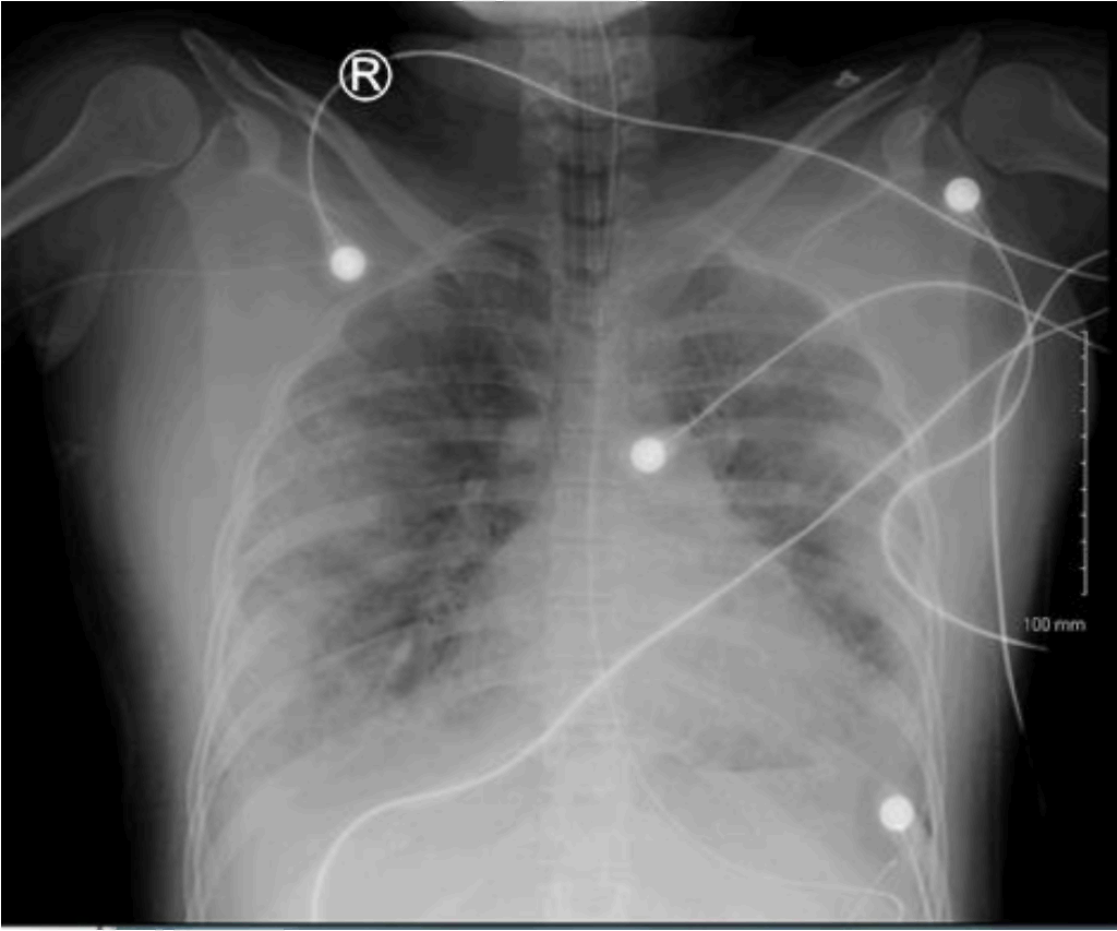
The physical examination on initial presentation revealed lung sounds clear to auscultation with no wheezes, rales or rhonchi, and cardiac sounds including regular rate and rhythm with no murmurs, rubs or gallops. The rest of the exam was largely unremarkable except for skin findings of petechiae on her right breast, abdomen, and left buttock. At the time of presentation, a chest radiograph was performed and showed fluffy opacities of the bilateral mid-to-lower lungs with a peripheral predominance (figure 1).
Figure 1.AP Chest Radiograph on Initial Presentation.
A chest computerized tomography (CT) without contrast on presentation also showed predominantly basilar and peripheral ground glass opacities/nodules bilaterally. A complete blood cell count taken at the same time had white blood cells within normal limits at 10.5 ×10⁹/L, but an absolute neutrophil count elevated at 9.1 × 10⁹/L. Arterial blood gas studies at that time were unremarkable. The patient was tested for influenza using rapid influenza diagnostic tests, infectious mononucleosis using mononuclear spot testing, and tuberculosis via acid-fast bacilli testing. All of these investigations were negative. The patient was also negative for HIV and Legionella infection. Another significant laboratory value initially was elevated Creactive protein (CRP) at 8.0 mg/L, demonstrating an inflammatory response.
The patient was admitted to the inpatient service for workup and on day two of her hospital course, experienced worsening dyspnea and tachypnea and requested intubation fearing further decline in her breathing status. She was then transferred to the ICU for intubation and mechanical ventilation. Collateral obtained from the patient's family following her intubation revealed she received silicone injections in her buttocks bilaterally for cosmetic purposes two days prior to symptom onset. This was a procedure she had reportedly received in the past as well. It was at this time that the medical team considered the likelihood of silicone embolism syndrome versus severe pneumonia. The patient was diagnosed with silicone embolism syndrome on day two of her hospital course.
Her status continued to deteriorate over 48 hours. Serial chest radiograph demonstrated bilateral fluffy opacities representing ARDS versus focal pneumonia, only mildly worsened compared to prior exam in the emergency department (Figure 2). On her fifth hospital day chest radiograph showed mild interval worsening of diffuse bilateral airspace disease, not worsened in bilateral upper lungs. There was a possible small bilateral pleural effusion and the findings were determined to be likely infectious or inflammatory. White blood cell counts trended over the course of her stay showed rapid increase; from 10.5 × 10⁹/L to 11.4 × 10⁹/L within her first 24 hours of presentation to a critical high of 21.8 × 10⁹/L on day five of her hospital course. Absolute neutrophil count also trended up from initial 9.1 × 10⁹/L on day one to a critical high of 17.71 × 10⁹/L on day five. CRP continued to increase from initial 8.0 mg/L, reaching 12.90 mg/L on day four of her hospital course.
Figure 2.AP Chest Radiograph 48-Hours After Admission.
On initial presentation this patient was treated with broad spectrum antibiotics based on her clinical deterioration and worsening opacities seen in her chest radiographs. Throughout her course she was treated with methylprednisolone initially at 40 mg IV every six hours. She continued to experience episodes of hemoptysis while intubated, contributing to a worsening anemia and necessitating a blood transfusion. The dose was eventually increased to 125 mg IV every six hours once she was transferred to the ICU. The patient was treated with prone positioning on her fifth hospital day while a transfer was arranged for her treatment with extracorporeal membrane oxygenation (ECMO) at a larger hospital.
After transfer and treatment with ECMO, the patient remained under the care of inpatient therapy at a hospital three months later. She recovered and was discharged from inpatient hospital care approximately four months following her presentation to the emergency department.
Silicone embolism syndrome is a condition that occurs when silicone injected for cosmetic or therapeutic purposes enters the systemic venous system and travels to the pulmonary vasculature. Proposed pathophysiology involves histiocytes and foreign-body giant cells migrating to the site of the micro emboli causing diffuse inflammation as well as alveolar hemorrhage.1 This diffuse inflammation develops rapidly as was demonstrated in this patient's laboratory CRP values, leukocyte and neutrophil counts, and serial radiograph readings. Evidence of alveolar hemorrhage is provided by the patient's history of hemoptysis prior to presentation and continued hemoptysis even while on mechanical ventilation. Future investigations into this syndrome may benefit from bronchoscopy and/or serial lung biopsies to demonstrate the progression of inflammatory cell migration in alveolar capillaries.
Similar to fat embolism syndrome (FES), silicone embolism syndrome patients may present with a characteristic petechial rash, hypoxemia, dyspnea, and tachypnea. In review of this patient's hospital course, it should be noted that the patient presented with characteristic petechiae. The presence of petechial rash and respiratory decline should have prompted the question of recent trauma in consideration of fat embolism syndrome at the very least. This patient's rare case should be considered as an example of the importance of detailed history taking. Future considerations for standard of care may involve the question of foreign body contact or injection when treating a patient with cutaneous manifestations and respiratory distress.
The complication rate of silicone injections in general are estimated to be about 1-2%,2 which includes silicone embolism to the lungs, making this syndrome particularly unusual. Given its similarity to fat embolism syndrome, guidelines for care and diagnosis are followed similarly. Both syndromes are clinical diagnoses and diagnoses of exclusion, therefore a wide differential diagnosis should be considered. The differential diagnosis for this patient included alveolar filling defects like pneumonia and ARDS, as mentioned after thoracic imaging studies. Given that the only treatment available for silicone embolism syndrome is supportive therapy and the diagnosis is clinical, a noninvasive diagnostic approach with chest imaging is appropriate. A full diagnostic workup of a patient with silicone embolism syndrome should include coagulation panels and serial complete blood cell counts to rule out disseminated intravascular coagulation (DIC) and other hypercoagulable states like inherited thrombophilia.
As in the case of FES, presence of the characteristic manifestations of hypoxemia, fever, and a petechial rash following injection of silicone is highly suggestive of the condition. When silicone embolism syndrome is suspected, chest radiography and/or CT is performed, as seen in the case described. When compared with other case studies of silicone embolism syndrome, the presence of ground glass opacities bilaterally on chest CT in this patient is characteristic and supports the diagnosis.3
Because at this time there is no definitive treatment for silicone embolism syndrome, management is supportive, as mentioned previously. The standard of care is followed in the patient described as she is treated with methylprednisolone, a corticosteroid. The effects of corticosteroids in this patient and others with ARDS include inhibition of hypoxic vasoconstriction, and reduction and modulation of inflammatory mediators. The goal of corticosteroid use in these patients is to control the inflammatory response while the syndrome resolves spontaneously.
The prognosis of silicone embolism syndrome is associated with rapid clinical deterioration. Management of this condition is primarily supportive, but prone ventilation is a recruitment maneuver that may improve oxygenation. The prone ventilation treatment approach is based on the theory of redistribution of the blood so that ventilation and perfusion is better matched. Through this mechanism regional changes in ventilation will ideally improve oxygenation. Benefits of prone positioning are supported by the results of the Proning Severe ARDS (PROSEVA) study, which revealed a significantly decreased 28-day and 90-day mortality rate in patients with severe ARDS who received early treatment with prone positioning sessions of at least 16 hours when compared to patients with severe ARDS who remained in the supine position.4 It should also be considered that the mortality benefits of prone positioning may be greatly associated with decrease in severe tissue strain and not simply increased oxygenation.5 The mortality rate of silicone embolism syndrome has been described to be approximately 25%.6
In summary, the cosmetic use of silicone via injection can result in entrance of the synthetic polymer into the systemic venous system and embolization to the pulmonary vasculature. These micro emboli can lead to diffuse alveolar hemorrhage associated with rapid respiratory decline and cutaneous manifestations similar to the presentation of fat embolism syndrome. Although this presentation is rare and diagnosis is not obvious, clinicians aware of the possible complications of such cosmetic procedures can potentially screen for risk factors with a detailed history and physical exam. Increased awareness of this rare condition can improve the prognosis for patients suffering from complications.
No acknowledgements to disclose.
The Authors have no funding, financial relationships or conflicts of interest to disclose.
Conceptualization: CM, and ZR. Methodology: CM, and ZR. Software: CM, and ZR. Validation: CM, and ZR. Formal Analysis: CM, and ZR. Data Curation: CM, and ZR. Investigation: CM, and ZR. Resources: CM, and ZR. Writing – Original Draft: CM, and ZR. Writing – Review & Editing: CM, and ZR. Visualization: CM, and ZR. Supervision: CM, and ZR. Project Administration: CM, and ZR. Funding Acquisition: CM, and ZR.
Arora A, Inaty H, Mukhopadhyay S, Abu-Hijleh M, Mehta AC. Chronic Pulmonary Silicone Embolism Related to Saline Breast Implants. Ann Am Thorac Soc. 2016 Jan;13(1):139-41.
Mendonca D, Leitao DS, Friend R, Epelboim J, Eiger G. An unusual case of pulmonary embolism. Respir Care. 2012 Aug;57(8):1345-7.
Dawn SK, Elicker BM, Leung JWT, Reddy GP, Gotway MB. The Silicone Syndrome. Clinical Pulmonary Medicine. 2006 Mar;13(2):146-7.
Guérin C, Reignier J, Richard JC, Beuret P, Gacouin A, Boulain T, et al. Prone positioning in severe acute respiratory distress syndrome. N Engl J Med. 2013 Jun 6;368(23):2l59-68.
Henderson WR, Griesdale DE, Dominelli P, Ronco JJ. Does prone positioning improve oxygenation and reduce mortality in patients with acute respiratory distress syndrome? Can Respir J. 2014 Jul-Aug;21(4):213-5.
Tobon-Trujillo M, Arias L, Jaramillo LF, Juyo A, Villaquiran-Torres C. Silicone Embolism Syndrome: A Case Series. Am J Respir Crit Care. Med. 2017:195:5564.
Carolyn Frances Molina, 1 Medical Student, OMS-III, Lake Erie College of Osteopathic Medicine - Bradenton Campus, LECOM Bradenton, FL, USA
Zachary Paul Retalis, 1 Medical Student, OMS-III, Lake Erie College of Osteopathic Medicine - Bradenton Campus, LECOM Bradenton, FL, USA
About the Author: Carolyn Molina is currently a fourth-year medical student of a four-year program at LECOM Bradenton in Bradenton, Florida, USA. She is pursuing a residency in internal medicine.
Correspondence: Carolyn Frances Molina. Address: 5000 Lakewood Ranch Blvd, Bradenton, FL 34211, USA. Email: CMolina91983@med.lecom.edu
Cite as: Molina CF, Retalis ZP. Pulmonary Embolism Secondary to Silicone Injection. Int J Med Students. 2019 May-Aug;7(2):41-44.
Copyright © 2019 Carolyn Frances Molina, Zachary Paul Retalis.
International Journal of Medical Students, VOLUME 7, NUMBER 2, August 2019