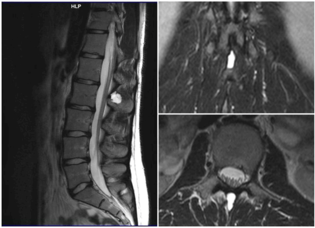
MRI scan. The MRI showed supernatants of the spinous process of the L2 vertebral body, an imprint located between the spinous processes of L1-L2 vertebrae.

Eliza (Eleni-Zacharoula) Georgiou1, Savvina Prapiadou1, Helen P. Kourea2
doi: http://dx.doi.org/10.5195/ijms.2019.407
Volume 7, Number 2: 33-37
Received 08 07 2019: Accepted 28 08 2019
ABSTRACT
Background:Aneurysmal bone cysts (ABC) are uncommon entities which cause expansile and destructive bone lesions and are characterized by reactive proliferation of connective tissue. They usually grow rapidly with hypervascularity. ABC's incidence on the spine is 1.5 in 10 million. Most cases present with pain of unexplained origin.
The Case:Presented in this paper is an ABC case in the spinous process of the L2 vertebra of a 20-year-old Creek female patient. The main symptom was persistent back pain, without neurological symptoms, of four years' duration. Treatment consisted of surgical curettage of the lesion. In this case report, we tried to describe not only the pathology of this disease but also the subsequent psychosocial symptoms that accompany it. We managed to accomplish that by exploiting the knowledge of an experienced pathologist, the help of the physicians responsible for this case, the interest of some sensitized medical students, and of course, the experience of the patient herself since the patient is also the lead author.
Conclusion:The focal point of this article is that even though ABCs might lead to excruciating pain, this pain can be alleviated with the proper treatment, especially if the communication between physician and patient is optimal.
Keywords: Aneurysmal Bone Cyst; Spine; Pain; Neoplasms (Source: MeSH-NLM)..
The aneurysmal bone cyst (ABC) is a benign tumor-like lesion that is described as "an expanding osteolytic lesion consisting of blood-filled spaces of variable size separated by connective tissue septae containing trabeculae or osteoid tissue and osteoclast-type giant cells.1 ABC's general incidence is 1.4 in a million, the median age is 13 years.2 Although benign, ABC can be locally aggressive. Its expansile nature can cause pain, swelling, deformity, disruption of growth plates or joint surfaces, neurological symptoms (depending on the location), and pathologic fracture.3
In this case report, the patient's identity is intentionally disclosed to the reader. As the patient, one of my main goals for writing this case report was to describe the evolution of my experience. More specifically, I wanted to highlight how this rare lesion shifted my perspective on chronic pain.
As medical students, we study extensively the pathology, anatomy and pathophysiology of every disease. However, we usually lack the ability to communicate with our patients and completely understand how they feel, mainly due to gaps in our educational system. This incident helped me see my future as a medical doctor from a different point of view. Having this rare lesion on my body, I realized just how devastating chronic pain can be. I understood that our role and responsibility as doctors is to not only to offer the appropriate disease related-treatment to our patients but also to truly listen to them, to show compassion and empathy, and to aid and guide them in the alleviation of the psychosocial symptoms they might be experiencing. In short, I recognized the importance of advocating for their well-being in whatever way we can and includes (and possibly begins with) rendering the correct diagnosis and using any and every resource available to us.
Key Points:
There are plenty of case studies for ABCs but none of them focus on the fact that this pain is capable of confining you in bed.
Additionally, they don't place enough emphasis on the fact that the pain can be alleviated with the proper treatment. ABCs might not be severe or risky, but it's terrifying to imagine living your life in constant pain.
The patient (16y, female, Greek) initially presented both on the orthopedic and athletic clinic with chronic back pain, intense spinal muscular spasms bilaterally and a clinical picture of slight left-curved scoliosis. Due to the patient's involvement in professional basketball, no further examination or imaging was considered necessary since the symptoms were directly attributed to intensive sport-related stress to the body. Non-steroidal Anti-Inflammatory drugs (NSAIDs) and muscle relaxants were prescribed. Physiotherapy sessions were also performed without causing any pain alleviation. One year after the onset of the pain, the patient was discouraged from continuing professional basketball and subsequently resigned. The pain and the symptoms persisted for 4 years.
Differential diagnosis at the time consisted of muscle strains, intervertebral disc herniation, trauma in the area and psychogenic pain.
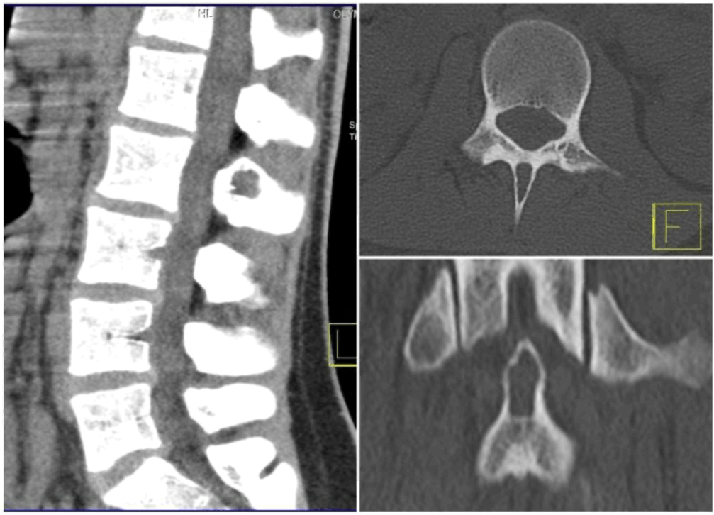
After 4 years from the initial onset of pain (age of patient 20y), the patient relapsed and became bedridden. A Magnetic Resonance Image (MRI) scan with contrast was performed. (Figure 1). MRI showed supernatants of the spinous process of the L2 vertebral body, an imprint located between the spinous processes of L1-L2 vertebrae. The lesion was clearly circumscribed and was not surrounded by swelling. It was measured with maximum dimensions of 1.2×1.6×0.6 cm. This alteration produced signal intensity similar to the cerebrospinal fluid (CSF) in T2 weighted sequences and signal intensity close to the bone marrow in T1 weighted sequences. It was considered necessary for the lesion to be further investigated by Computerized Tomography (CT) scanning focusing at L2 to document the relation of this lesion to the spinous process. (Figure 2).
Figure 1.MRI scan. The MRI showed supernatants of the spinous process of the L2 vertebral body, an imprint located between the spinous processes of L1-L2 vertebrae.

CT scan. The altered lesion was elucidated in this test as it is located in the spinous process of L2.

The altered lesion was elucidated in this test as it was located in the spinous process of L2. The upper part of the lesion showed a labyrinthine edge and had broken mildly the bone. It had maximum dimensions in the sagittal level of 1,6 × 1,4 cm. The lesion had clear and smooth confines, and mildly extended to the bone without breaking the cortex. No other visible bone alteration was apparent from the study of the bone section shown. Based on the above imaging findings, the lesion had mainly benign characteristics. The protagonist of differential diagnosis was an aneurysmal bone cyst. The chance of giant cell tumor was considered low.
The opinions regarding treatment were discordant. Paracetamol and muscle relaxants (3 pills of 1000mg paracetamol & orphenadrine per day) were prescribed for 6 months without pain remission. The patient was bedridden for that time interval and consequently she could not accomplish her daily obligations. She was not able to attend the lectures and workshops of her medical school which caused her academic performance to decline. At the same time, she was isolated in her home because she could not cope with the physical requirements of social events.
After that period of time, surgical removal of the L2 spinous process and cyst wall was performed through a 4cm incision. The resected material consisted of multiple tissue fragments measuring 4×3.5×0.7cm, including pieces of bone, striated muscle and fibrofatty tissue. NSAIDs were given for 2 weeks after surgery. The patient avoided the sitting position, torsional movements and lifting weights for 2 months.
The biopsy material included mainly cancellous bone containing hematopoietic bone marrow, skeletal muscle and fibrofatty tissue fragments. Focally, among the bone spicules and marrow elements there were small fragments of a cystic lesion, lined by flattened endothelium, containing few red blood cells. These findings were consistent with ABC.
The recovery included the empowerment of the muscles that support the spine. NSAIDs were given for 2 weeks after the surgery and an orthopedic band was used to support spinal muscles. One year after the surgery, there are episodes of bilateral numbness in the legs and back pain in the area of the posterior iliac process usually lasting a week. These symptoms may be attributed to the operation-related development of connective tissue that presses the spinal cord.
However, the patient is capable of lifting weights, of engaging in sports and of living without the constant need for NSAIDs. The remission of the pain makes her capable of not only improve her physical health, but also her psychosocial life because she can make long term plans without the fear of a relapse and the excruciating pain it is accompanied by.
The annual incidence of primary aneurysmal bone cyst was 0.14 per 100.000 individuals; however, the true incidence is difficult to calculate because of the existence of spontaneous regression and clinically silent cases. In a published review of 897 cases of ABC, the following rates of occurrence were reported ( Table 1) and as we stated on the abstract, the occurrence of spine ABCs is 1.5 in 10 million3.
Table 1.Rates of occurrence.
| Sites of involvement | Frequency |
|---|---|
| Tibia | 17.5% |
| Femur | 15.9% |
| Vertebra | 11.2% |
| Pelvis | 11.6% |
| Humerus | 9.1% |
| Fibula | 7.3% |
| Foot | 6.3% |
| Hand | 4.7% |
| Ulna | 3.8% |
| Radius | 3.1% |
| Other | 9.2% |
In this case, due to limited resources, we were unable to investigate the etiology and pathophysiology of ABC. However, through a literature review we found that true etiology of ABCs is as of yet unknown. Most investigators believe that ABCs are the result of a vascular malformation within the bone; however, the ultimate cause of the malformation is a topic of controversy. There are three commonly proposed theories (Table 2).4
Table 2.Etiology theories of ABCs.
| Etiology theories of ABCs | ||
| ABCs may be caused by a reaction secondary to another bone lesion (ABCs in the presence of another lesion are called secondary ABCs, and treatment of these ABCs is based on what is appropriate for the underlying tumor). | ABCs may arise de novo; those that arise without evidence of another lesion are classified as primary ABCs. | ABCs may arise in an area of previous trauma. |
The true pathophysiology of ABCs is also unknown.5 Different theories about several vascular malformations exist; these include arteriovenous fistulas and venous blockage. The vascular lesions then cause increased pressure, expansion, erosion, and reabsorption of the surrounding bone. The malformation is also believed to cause local hemorrhage that initiates the formation of reactive osteolytic tissue. Findings from a study in which manometric pressures within the ABCs were measured support the theory of altered hemodynamics. Most primary ABCs demonstrate a t(16;17)(q22;p13) fusion of the TRE17/CDH11-USP6 oncogene. This fusion leads to increased cellular cadherin-11 activity that seems to arrest osteoblastic maturation in a more primitive state. This process may be the neoplastic driving force behind primary ABCs as opposed to secondary ABCs, which seem to as a reaction to underlying disease process.6,7
Patients with an aneurysmal bone cyst (ABC) usually present with pain, swelling, a mass at times rapidly enlarging, or pathologic fracture in the affected area, or even a combination of the above. The symptoms are usually present for several weeks to months before the diagnosis is rendered. Neurologic symptoms associated with ABCs in the spine may develop secondary to pressure or tension of nerves over the lesion. Pathologic fracture occurs in about 8% of ABCs, but the incidence may be as high as 21 % in ABCs that have spinal involvement.
Aneurysmal bone cysts (ABCs) are generally treated surgically8 which was the approach that was followed by our patient. Rarely, asymptomatic ABCs may be seen in which there is clinically insignificant destruction of bone. In such cases, close monitoring alone of the lesion may be indicated because of the evidence that some ABCs spontaneously resolve. When a patient is monitored in this manner, the diagnosis must be certain, and the lesion should not be increasing in size.
Some anatomic locations may be difficult to access surgically. If this situation is encountered, other methods of treatment, such as intralesional injection and selective arterial embolization, may be successful. In the future, advances in osteoinductive materials (eg, genetically engineered bone morphogenic protein) may offer a less invasive treatment of ABC.8,9
Throughout this case report we described several of the psychosocial symptoms of the patient which were a direct result of the chronic pain caused by the ABC. Additionally, we thoroughly explained the biometric aspects of ABC. The emphasis that was placed on both aspects of the patient's condition is meant to highlight the importance of a biopsychological approach of every patient.
The biopsychosocial approach was developed by Drs. George Engel and John Romano in 1977. While traditional biomedical models of clinical medicine focus on pathophysiology and other biological approaches to diseases, the biopsychosocial approach emphasizes the importance of understanding human health and illness in their fullest contexts. The biopsychosocial approach systematically considers biological, psychological, and social factors and their complex interactions in understanding health, illness, and healthcare delivery.11
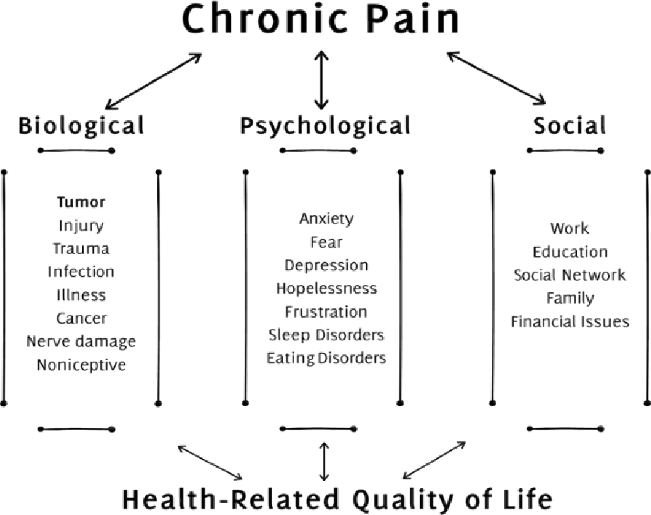
Chronic pain affects a large proportion of the population, imposing significant individual distress and a considerable burden on society, yet treatment is not always instituted and/or adequate. Comprehensive multidisciplinary management based on the biopsychosocial model of pain has been shown to be clinically effective and cost-efficient, but is not widely available (Figure 3).
Figure 3.Biopsychosocial model of chronic pain and consequences on the quality of life12

Many of the issues relating to physicians could be addressed by improving medical training, both at undergraduate and postgraduate levels – for example, by making pain medicine a compulsory core subject of the undergraduate medical curriculum. This would improve physician/patient communication, increase the use of standardized pain assessment tools, and allow more patients to participate in treatment decisions. Patient care would also benefit from improved training for other multidisciplinary team members; for example, nurses could provide counseling and follow-up support, psychologists offer coping skills training, and physiotherapists could have a greater role in rehabilitation. Equally important measures include the widespread adoption of a patient-centered approach, chronic pain being recognized as a disease in its own right, and the development of universal guidelines for managing chronic, non-cancerous pain.13
This case report is written in memory of Dimitrios Konstantinou.
We want to truly thank the neurosurgeon Dimitrios Konstantinou, who performed the surgical curettage of this lesion but unfortunately passed away a few weeks later. He was an inspiration and a role model not only for his medical skills but also for his ability to empathize with his patients.
The Authors have no funding, financial relationships or conflicts of interest to disclose.
Conceptualization: E.Z.C. Writing – Original Draft: E.Z.C. Writing – Review & Editing: S.P., and H.P.K. Visualization: E.Z.G. Supervision: H.P.K. Project Administration: E.Z.C.
Schajowicz F. Aneurysmal bone cyst. Histologic Typing of Bone Tumours. Berlin: Springer-Verlag; 1992. 37
.Leithner A, Windhager R, Lang S, et al. Aneurysmal bone cyst. A population based epidemiologic study and literature review. Clin Orthop Relat Res. 1999 lun. (363)1176-9.
Burch S, Hu S, Berven S. Aneurysmal bone cysts of the spine. Neurosurg Clin N Am. 2008 Jan. 19(1)141-7.
Kransdorf MJ, Sweet DE. Aneurysmal bone cyst: concept, controversy, clinical presentation, and imaging. AJR Am J Roentgenol. 1995 Mar. 164(3):573-80.
Cottalorda J, Bourelle S. Modern concepts of primary aneurysmal bone cyst. Arch Orthop Trauma Surg. 2007 Feb. I27(2):105-14.
Lau AW, Pringle LM, Quick L, Riquelme DN, Ye Y, Oliveira AM, et al. TRE17/ubiquitin-specific protease 6 (USP6) oncogene translocated in aneurysmal bone cyst blocks osteoblastic maturation via an autocrine mechanism involving bone morphogenetic protein dysregulation. J Biol Chem. 2010 Nov 19. 285(47):37111-20.
Guseva NV, Jaber 0, Tanas MR, Stence AA, Sompallae R, Schade J, et al. Anchored multiplex PCR for targeted next-generation sequencing reveals recurrent and novel USP6 fusions and upregulation of USP6 expression in aneurysmal bone cyst. Genes Chromosomes Cancer. 2017 Apr. 56 (4)1266-277.
Park HY, Yang SK, Sheppard WL, Hegde V, Zoller SD, Nelson SD, et al. Current management of aneurysmal bone cysts. Curr Rev Musculoskelet Med. 2016 Dec. 9 (4):435-444.
Schreuder HW, Veth RP, Pruszczynski M, et al. Aneurysmal bone cysts treated by curettage, cryotherapy and bone grafting. J Bone Joint Surg Br. 1997 Jan. 79(0):20-5.
Brastianos P, Gokaslan Z, McCarthy EF. Aneurysmal bone cysts of the sacrum: a report of ten cases and review of the literature. Iowa Orthop J. 2009;29:74-8
Engel GL: The clinical application of the biopsychosocial model. Am J Psychiatry 1980;137:535-544
Maria Dueñas, Begoña Ojeda, Alejandro Salazar, Juan Antonio Mico, and Inmaculada Failde A review of chronic pain impact on patients, their social environment and the health care system June 2016 Journal of Pain Research Volume 9(lssue 0:457-467
Kress HG, Aldington D, Alon E, Coaccioli S, Collett B, Coluzzi F et al. A holistic approach to chronic pain management that involves all stakeholders: change is needed. Curr Med Res Opin. 2015;31(9):1743-54
Eliza (Eleni-Zacharoula) Georgiou, 1 Medical Student, University of Patras, Greece
Savvina Prapiadou, 1 Medical Student, University of Patras, Greece
Helen P. Kourea, 2 Associate Professor of Pathology, University of Patras, Greece
Mihnea-Alexandru Găman, Editor
About the Author: Eliza (Eleni-Zacharoula) Georgiou is a 4th year medical student at the University of Patras, Greece. Her biggest passion is Mental Health with somatic symptoms and disorders.
Correspondence: Eliza (Eleni-Zacharoula) Georgiou. Address: Medical School, University of Patras Greece, Πανεπιστηιούπoλη Πατρών 265 04, Greece. Email: elizageo8@gmail.com
Cite as: Georgiou E, Prapiadou S, Kourea HP. Spine ABC, A Multidimensional Case Report from A to Z: Aneurysmal Bone Cyst of the Spine. In memory of Dimitrios Konstantinou. Int J Med Students. 2019 May-Aug;7(2):33-7.
Copyright © 2019 Eliza (Eleni-Zacharoula) Georgiou, Savvina Prapiadou, Helen P. Kourea
International Journal of Medical Students, VOLUME 7, NUMBER 2, August 2019