Case Report
Early Multifocal Recurrence of Incidentally Discovered Pulmonary Epithelioid Hemangioendothelioma Following Complete Surgical Resection
Lilia Crew1, Jeanine Chiaffarano2, Brian Mott3
doi: https://doi.org/10.5195/ijms.2026.4334
Volume 14, Number 2: 107-111
Received 07 01 2026; Rev-request 26 03 2026; Rev-request 26 05 2026; Rev-recd 04 04 2026; Rev-recd 30 05 2026; Accepted 27 07 2026
ABSTRACT
Background:
Pulmonary epithelioid hemangioendothelioma is a rare, highly vascularized tumor with variable clinical presentation including recurrence and metastases. The treatment is complicated by the lack of standardized guidelines along with differing responses to trialed treatments, leaving patient prognosis unpredictable. Although often indolent, recurrence patterns remain poorly characterized in literature, specifically in patients with initially localized disease.
Thes Case:
We report a case of a 77-year-old man in whom a left, 21 mm lower-lobe pulmonary nodule was incidentally discovered during pre-coronary artery bypass graft evaluation. A concurrent wedge resection was performed with histopathology confirming epithelioid hemangioendothelioma, with negative margins. The patient opted for surveillance due to asymptomatic presentation. A year later, a recurrence with a right lower-lobe lesion, 10 × 7 mm, and numerous scattered lesions were identified and resected, both consistent with metastatic/recurrent epithelioid hemangioendothelioma. Histology from the initial and recurrent resections showed epithelioid nests and necrosis with immunopositivity for CD31, CD34, ERG, CAMTA1, and vimentin.
Conclusion:
This case emphasizes the importance of long-term surveillance in pulmonary epithelioid hemangioendothelioma due to the potential for early or unexpected recurrence as well as the need for more research to investigate a standardized, yet customizable, approach for better patient outcomes.
Keywords: Epithelioid Hemangioendothelioma, Lung Neoplasms, Vascular Neoplasms, Thoracic Surgery, Neoplasm Recurrence
Introduction
Epithelioid hemangioendothelioma (EHE) is a rare vascular neoplasm that can arise in multiple organs, such as the lung, liver, and bone, with variable metastatic behavior.1,2 Epidemiologically, EHE has an estimated incidence of less than one case per million population.3,4 It typically occurs in adults between their third and fifth decades, with a slight predominance in females.
Pulmonary EHE accounts for less than 1% of vascular tumors and is often an incidental finding on imaging, as 50-76% of patients with EHE are asymptomatic.5 In older individuals, pulmonary nodules typically raise suspicion for primary lung carcinomas or metastatic disease, making the diagnosis of EHE particularly challenging. Additionally, the unpredictable course of EHE can further add to its misdiagnosis or late diagnosis, delaying treatment.
Histologically, EHE is characterized by epithelioid endothelial cells within a myxohyaline stroma with varying mitotic activity.6 Immunohistochemical staining for typical endothelial/vascular markers such as CD31, CD34, and FLI-1 is essential in confirming the diagnosis and distinguishing the lesion from epithelial and mesenchymal malignancies.
EHE ranges from indolent to aggressive with metastatic potential. 7,8 The mainstay of treatment is surgical resection; however, multifocal lesions make this unfeasible in advanced cases, resulting in the need for chemotherapy. Previous studies have shown that the 5-year disease-specific survival for EHE is 81%, sharply contrasting the 50% mortality rate at 1 year for soft-tissue angiosarcomas.8
Prognosis varies widely and is influenced by factors such as pleural effusion, presence of symptoms at diagnosis, and extrapulmonary metastasis–all of which are associated with poorer survival outcomes.3,9
Multifocal recurrence within one year of resection is rarely reported in pulmonary EHE. To contextualize this case within existing literature, a targeted search of PubMed and Google Scholar using the terms ‘pulmonary epithelioid hemangioendothelioma”, “pulmonary EHE recurrence,’ and ‘pulmonary EHE resection’ identified only a small number of case-level reports describing recurrence within this timeframe following negative-margin resection. The case discussed herein adds to the limited body of evidence and highlights the need for long-term surveillance post-resection.
The Case
Patient Presentation
The patient was a 77-year-old Caucasian male, a former smoker with fewer than 10 pack-years, having quit 16 years prior. He had no known occupational exposures or family history of malignancy. His past medical history included hypertension, hyperlipidemia, type 2 diabetes mellitus, osteoarthritis, spine radiculopathies, coronary atherosclerosis, and a cavernous hemangioma of the spine. He reported minor alcohol usage and used a marijuana tincture for pain related to the spinal hemangioma. He was not on cytotoxic agents or steroids despite his multiple comorbidities. The patient denied chest pain, cough, hemoptysis, fever, and chills.
Figure 1.Epithelioid Hemangioendothelioma — Serial Axial Chest CT and PET/CT Imaging
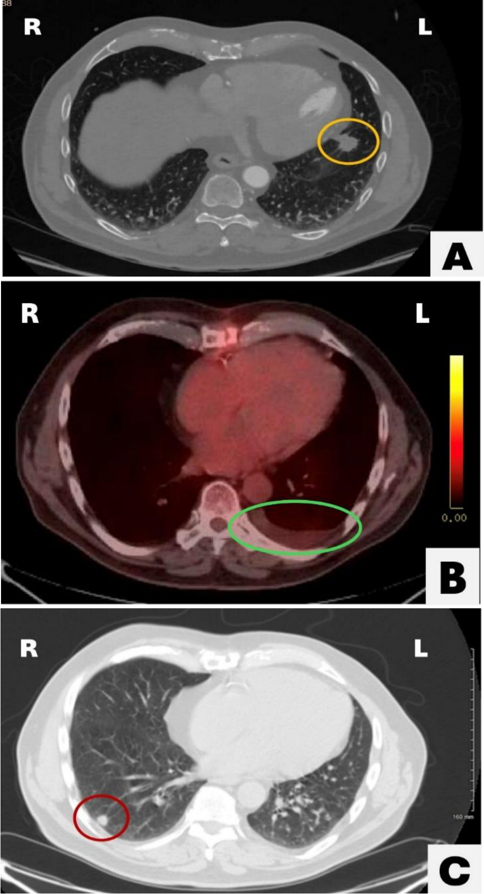
Legend: (A) Pre-operative axial CT demonstrating a left lower lobe pulmonary nodule (orange circle), representing the primary resected lesion. (B) Axial PET/CT performed 3 months post-operatively demonstrating a residual left pleural effusion (green circle) without evidence of metabolically active recurrence or residual nodule(s). (C) CT component of axial PET-CT at 12 months post-operatively demonstrating bilateral pulmonary nodules, including a right lower lobe subpleural nodule (red circle, largest approximately 10 × 7 mm). The CT portion is displayed as non-avid lesions are better characterized by CT.
During cardiac evaluation for coronary artery disease, pre-operative CT chest imaging identified a 21 mm spiculated left lower lobe pulmonary mass. Subsequent PET-CT demonstrated moderate radiotracer uptake at this site (Standard Uptake Value-SUV 2.3, Figure 1A), raising suspicion for primary lung malignancy. Routine preoperative laboratory workup, including complete blood count (CBC) and comprehensive metabolic panel (CMP), was unremarkable. Further targeted workup, including inflammatory markers (CRP, ESR) and sputum AFB, was not obtained, as the imaging characteristics and clinical presentation were not suggestive of an infectious etiology.
One month after pre-surgical screening, the decision was made to perform a three-vessel coronary artery bypass grafting (CABG) with a concurrent generous wedge resection due to peripheral location and localization of the tumor, while minimizing the need for multiple operations (Table 1). Pathology from the resected lesion revealed epithelioid hemangioendothelioma with negative surgical margins, representing localized, single-lesion pulmonary disease. The patient expressed a preference for surveillance over systemic therapy, citing his stable condition and desire to avoid treatment-related morbidity. One-month postoperatively, a small left pleural effusion was identified on imaging but was not drained due to its minimal size.
Figure 2.Histology Hematoxylin and Eosin Stain, 10x
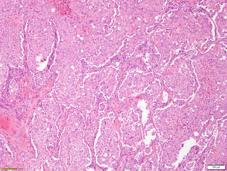
Legend: (Wedge resection of lung showing solid nests of tumor cells expanding airspaces. Rhabdoid features and foci of necrosis are noted. This morphology, along with subsequent immunostaining, is consistent with epithelioid hemangioendothelioma.
At 3 months post-operatively, surveillance PET-CT demonstrated a decreased left pleural effusion, consistent with post-operative changes, with no nodules or suspicious metabolic uptake identified (Figure 1B). Following this, the patient opted for yearly surveillance imaging; he was counseled that earlier imaging would be warranted should his clinical status change. One year after his initial surgery, PET-CT surveillance imaging performed by oncology identified a 10 × 7 mm right lower lobe (RLL) nodule with low metabolic activity (SUV 0.9), along with multiple non-avid bilateral pulmonary nodules (Figure 1C). The patient again elected for surveillance over intervention given low-avidity nodules and continued asymptomatic status. A follow-up scan four months later showed the RLL nodule increased to 14 mm with moderate metabolic activity (SUV 2.6); additional non-avid bilateral pulmonary nodules were also noted. No metabolically active mediastinal lymphadenopathy was observed. The patient continually denied B symptoms, cough, and hemoptysis. On exam, no cervical lymphadenopathy was present. Pulmonary function tests were obtained preoperatively and values deemed adequate for surgical resection, at which point the patient decided to proceed with surgery. Due to the peripheral occurrence of the RLL lesion, a repeat robotic surgical wedge resection was performed, and pathology confirmed recurrent EHE. During this procedure, multiple pleural-based whitish nodules, each measuring approximately 1–2 mm, were observed scattered across the lung surface. A biopsied nodule confirmed EHE, supporting the presence of multifocal bilateral pulmonary disease. The postoperative course was uncomplicated.
Table 1.Clinical Timeline: Summary of key diagnostic, surgical, and surveillance events throughout the patient’s disease course.
Outcome
At two months following the second resection, the patient remained clinically stable with no new symptoms and postoperative laboratory studies were unremarkable. No systemic therapies had been administered. The patient continued under active oncologic surveillance with serial imaging.
Histology
The hematoxylin and eosin-stained sections, from the initial and most recent sampling, show an epithelioid neoplasm with solid nests expanding airspaces (Figure 2). Foci of tumor necrosis and rhabdoid features are noted. The tumor cells are positive for CD34, CD31, ERG, CAMTA1, and vimentin. They are negative for pancytokeratins, TTF-1, p40, S100, SOX10, HMB45, NKX3.1, PAX8, CD45 and calretinin. Overall, morphology and immunophenotype are consistent with epithelioid hemangioendothelioma with rhabdoid features. Molecular testing was not performed in this patient.
This case report was prepared in compliance with the CARE (Case Report) guidelines, informed written patient consent was obtained, and the case was deemed IRB exempt due to deidentification:
Discussion
Pulmonary nodules, particularly in older individuals with comorbidities, warrant a more extensive and thorough workup with a broad differential diagnosis. The initial considerations should include primary lung carcinoma, metastatic disease, and granulomatous infections such as tuberculosis or fungal causes. For this patient, the initial lesion with spiculated morphology and an SUV of 2.3 raised suspicion of malignancy, prompting biopsy. Histology showed an epithelioid neoplasm forming solid nests with focal necrosis and rhabdoid features, findings associated with more aggressive behavior and higher mortality in vascular neoplasms.9 In the case of EHE, immunohistochemistry is critical in confirming the diagnosis, demonstrated by the presence of endothelial/vascular marker CD34 positivity in this patient’s lesion.9 The negative pancytokeratin, TTF-1, and p40 excluded epithelial malignancies such as adenocarcinoma or squamous cell carcinoma.10
Diagnosis of EHE requires histopathologic evaluation with positive endothelial markers such as CD31, CD34, or FLI-1.9 It often occurs incidentally, as in this case, detected during pre-operative imaging for the CABG. Despite complete resection and negative margins of the 21 mm lesion, the patient developed a new right lower lobe lesion and scattered nodules within one year. In the registry analysis by Lau et al., progression from localized to uncontained or multifocal disease typically occurred over several years, with a median time to progression of 2.0 years.4 The Surveillance, Epidemiology, and End Result (SEER) database studied by Liu et al., further highlights the rarity of respiratory EHE and its association with worse overall survival in comparison to other anatomic locations, underscoring the clinical significance of early multifocal recurrence in this setting.3 This pattern raises the question of whether these lesions represent a single multifocal disease or independent occurrences. This case contributes to the evolving understanding of pulmonary EHE behavior.7,11
Of note, a study in 2011 identified recurrent WWTR1-CAMTA1 and YAP1-TFE gene fusions as characteristic molecular alterations in EHE. These fusions can be detected by FISH or RNA sequencing. CAMTA1 immunohistochemical positivity serves as a reliable surrogate marker for the WWTR1-CAMTA1 fusion, though it does not constitute molecular confirmation. In this case, molecular testing was not performed, which represents a limitation in the characterization of the disease, given the increasing clinical relevance of molecular classification in EHE.12
In the absence of standardized guidelines, management varies from surgical resection to systemic therapy for multifocal lesions or symptomatic cases. Agents such as interferon-alpha, bevacizumab, and mTOR inhibitors (e.g. sirolimus) have been used with variable success in advanced disease.13-15 In this case, the patient remained asymptomatic through both the initial diagnosis and recurrence, making an informed decision to opt for continued surveillance in both instances. Systemic therapy was deferred due to his stable condition and desire to avoid treatment-related morbidities, reflecting a shared decision-making approach that prioritized quality of life.
This case contributes to the growing understanding of EHE, specifically pulmonary EHE, by documenting early multifocal recurrence following complete resection in an elderly individual. The rapid recurrence observed in this case underscores the importance of long-term surveillance, even in patients with initially localized disease. In the absence of EHE-specific surveillance guidelines, structured imaging intervals like those used in post-treatment lung cancer follow-up may provide a potential framework for discussion, though this suggestion is hypothesis-generating rather than practice-informing and requires prospective validation. As more cases are reported, registries and longitudinal studies will be essential in defining prognostic factors and facilitating evidence-based management of this neoplasm. Documenting such early recurrence helps refine patients’ and providers’ expectations for surveillance intervals.
This report is limited by its single-patient nature, the rarity of pulmonary EHE, the patient's decisions regarding systemic treatment and routine surveillance restricting generalizability, the absence of molecular fusion testing, the unavailability of immunohistochemical staining images for inclusion, and the absence of targeted infectious workup including inflammatory markers (CRP, ESR, etc.) and sputum AFB studies, as these were not clinically pursued given the presentation.
Learning Points
Solitary lung nodules typically point clinicians towards primary lung cancers, metastasis or benign conditions like hamartomas or infections. A high suspicion for rare tumors, such as EHE, should be maintained when the clinical picture does not align with common etiologies. The use of immunohistochemistry is integral in establishing this diagnosis. Early tissue acquisition is important to enable timely multidisciplinary discussion, recognizing that evidence-based therapeutic standards for pulmonary EHE remain limited.
Conclusion
This case highlights the importance of long-term surveillance in patients with pulmonary EHE, even with localized initial lesions. The absence of standardized treatment guidelines shows that more research is required to allow patients and providers to approach this disease with greater direction and confidence.
Conflict of Interest Statement & Funding
The Authors have no funding, financial relationships or conflicts of interest to disclose.
Author Contributions
Conceptualization: L.C., B.M, Data Curation: L.C., B.M., J.C., Investigation: L.C., B.M., Supervision: B.M., J.C., Validation: B.M. ,J.C, Writing - Original Draft: L.C., Writing – review & editing: L.C., B.M., J.C.
References
1. Mardani P, Hashempour Z, Shahriarirad R, Amirian A. Epithelioid hemangioendothelioma prognosis and outcome: report of two challenging cases. J Cardiothorac Surg. 2024;19(1). doi: 10.1186/s13019-024-03338-x
2. Perea LA, Acebes EO, Mata NM, Pérez RS, Castel EV, Aragoneses FG. Hemangioendotelioma epiteloide pulmonar. Arch Bronconeumol. 2009;45(9):466–8.
3. Liu Z, He S. Epithelioid hemangioendothelioma: incidence, mortality, prognostic factors, and survival analysis using the SEER database. J Oncol. 2022;2022:1–10.
4. Lau K, Massad M, Pollak C, Rubin C, Yeh J, Wang J, et al. Clinical patterns and outcome in epithelioid hemangioendothelioma with or without pulmonary involvement. Chest. 2011;140(5):1312–8.
5. Sardaro A, Bardoscia L, Petruzzelli MF, Portaluri M. Pulmonary epithelioid hemangioendothelioma: an overview. World J Surg Oncol. 2014;12:259.
6. Jang JK, Thomas R, Braschi-Amirfarzan M, Jagannathan JP. A review of the spectrum of imaging manifestations of epithelioid hemangioendothelioma. AJR Am J Roentgenol. 2020;215(5):1290–8.
7. Lytle M, Bali SD, Galili Y, Bednov B, Murillo Alvarez RM, Carlan SJ, et al. Epithelioid hemangioendothelioma: a rare case of an aggressive vascular malignancy. Am J Case Rep. 2019;20:864–7. doi: 10.12659/AJCR.915874
8. Shiba S, Imaoka H, Shioji K, Suzuki E, Horiguchi S, Terashima T, et al. Clinical characteristics of Japanese patients with epithelioid hemangioendothelioma: a multicenter retrospective study. BMC Cancer. 2018;18(1):993.
9. Flucke U, Vogels RJ, de Saint Aubain Somerhausen N, Creytens DH, Riedl RG, van Gorp JM, et al. Epithelioid hemangioendothelioma: clinicopathologic, immunohistochemical, and molecular genetic analysis of 39 cases. Diagn Pathol. 2014 Jul 1;9:131.
10. Inamura K. Update on immunohistochemistry for the diagnosis of lung cancer. Cancers (Basel). 2018;10(3):72. doi: 10.3390/cancers10030072
11. Bagan P, Hassan M, Barthes FLP, Peyrard S, Souilamas R, Danel C, et al. Prognostic factors and surgical indications of pulmonary epithelioid hemangioendothelioma: a review of the literature. Ann Thorac Surg. 2006;82(6):2010–3.
12. Errani C, Zhang L, Sung YS, Hajdu M, Singer S, Maki RG, et al. A novel WWTR1 CAMTA1 gene fusion is a consistent abnormality in epithelioid hemangioendothelioma of different anatomic sites. Genes Chromosomes Cancer. 2011;50(8):644–53.
13. Tang B, Zhao S, Wang J, Xiang M, Dai J, Liu Q. Pulmonary epithelioid haemangioendothelioma: a comprehensive review of clinical and molecular advances. Clinics. 2025;80:100789.
14. Stacchiotti S, Simeone N, Lo Vullo S, Baldi GG, Brunello A, Vincenzi B, et al. Activity of sirolimus in patients with progressive epithelioid hemangioendothelioma: a case series analysis within the Italian Rare Cancer Network. Cancer. 2020;127(4):569–76.
15. Liu X, Zhou R, Si S, Liu L, Yang S, Han D, et al. Successful treatment with combined interferon alpha 2b and anlotinib in advanced hepatic epithelioid hemangioendothelioma: a case report. Front Med. 2022;9.
Lilia Crew, 1 Touro College of Osteopathic Medicine, USA.
Jeanine Chiaffarano, 2 DO, Pathology Associates of Northeast Pennsylvania, USA.
Brian Mott, 3 MD, FACS, FRCSC.
About the Author: Lilia Crew is a third-year medical student studying at Touro College of Osteopathic Medicine.
Correspondence: Lilia Crew. Address: 230 West 125th Street, New York, NY 10027. Email: lcrew@student.touro.edu
Process: Peer-reviewed
Cite as Crew L, Chiaffarano J, Mott B. Case Report: Early Multifocal Recurrence of Incidentally Discovered Pulmonary Epithelioid Hemangioendothelioma Following Complete Surgical Resection. Int J Med Stud. 2026 Apr-Jun;14(2): 107-111.
© 2026 Lilia Crew, Jeanine Chiaffarano, Brian Mott
This work is licensed under a Creative Commons Attribution 4.0 International License
International Journal of Medical Students, VOLUME 14, NUMBER 2, November 2026