Case Report
A Case Report Looking at an Incidental Finding of a Partial Anomalous Pulmonary Venous
Connection Using Magnetic Resonance Angiography
Iqra Qamar1, Mohammad Waleed2
doi: http://dx.doi.org/10.5195/ijms.2018.5
Volume 6, Number 1: 28-30
Received 23 12 2017:
Accepted 23 03 2018
ABSTRACT
Background
Partial anomalous pulmonary venous connection (PAPVC) is a rare congenital defect
where one or more of the pulmonary veins drain to the right atrium or its tributaries.
This can cause a left to right shunt (LTRS) which may need surgical management if
symptomatic.
Results
A 58-year-old female presented in emergency with non-pleuritic, central chest pain.
She denied breathlessness and fever. Her past medical history included: non-ST elevated
myocardial infarction (2013), anti-phospholipid syndrome, asthma, pulmonary embolism
(PE) and deep vein thrombosis many years prior. Differential diagnoses included myocardial
infarction, PE and unstable angina. Pulmonary angiography ruled-out PE. Coronary angiography
did not show obstructive coronary artery disease. On cardiac MRA an incidental finding
of PAPVC was found (left upper pulmonary vein drained into brachiocephalic vein) creating
a LTRS which wasn't substantial (Qp/Qs <1.5). Despite being asymptomatic, this patient
was followed-up of her incidental finding, and if symptoms developed she would be
considered for PAPVC correction surgery.
Conclusion
This case demonstrated the incidental finding of PAPVC in an otherwise asymptomatic
patient. The decision was not for surgery as the patient was not symptomatic from
the PAPVC and for follow-up. Previous studies showed PAPVC patients developing symptoms
of LTRS are candidates for operation. Physicians should be aware of rare anomalies
like PAPVC, their complications if unresolved, and should follow-up these patients
closely in case of worsening of the LTRS, in which case surgical correction may be
possible. Overall, PAPVC i) can be overlooked ii) diagnosis requires multimodality
imaging iii) constant follow up and iv) may be operable.
Keywords:
Anomalous Pulmonary Venous Return;
Cardiac Magnetic Resonance Angiogram;
Pulmonary Vein;
Left To Right Shunt;
Superior Vena Cava (Source: MeSH-NLM)..
Introduction
Partial anomalous pulmonary venous connection (PAPVC) is a congenital anomaly in which
one or more of the pulmonary veins drain to the right atrium or its tributaries, instead
of the left atrium, causing a left to right shunt (LTRS).1 The prevalence of incidental PAPVC is 0.1-0.7% and connection of the left upper pulmonary
vein to the superior vena cava (SVC) is rare.1,2-4 PAPVC can have associated congenital anomalies like atrial septal defect and has
been reported to be syndromic (e.g. Turner syndrome) in up to 25% of patients.5-6 In adults, PAPVC can be silent or present with its complications like dyspnea secondary
to LTRS. If left undiagnosed, complications such as right ventricular hypertrophy
can occur secondary to pulmonary hypertension. Ultimately, this can lead to right
sided (RS) overload and RS heart failure.7, 8
The Case
A 58-year-old female presented to the emergency department with non-pleuritic, central
chest pain. She denied any breathlessness and did not report feeling febrile. She
had a past medical history significant for a non-ST elevated myocardial infarction
(non-STEMI) in 2013 with a drug eluting stent to the syndrome (APS), asthma, pulmonary
embolism (PE) and deep vein thrombosis (DVT) many years prior. Furthermore, she was
on aspirin due to APS, but no current anticoagulation. Therefore, differential diagnoses
included myocardial infarction (Ml), PE and unstable angina of which pulmonary angiography
ruled-out PE.
Investigation findings showed T wave inversion in leads V2-V5 on EKG and an elevated
Troponin T (532 ng/L) confirming an NSTEMI. Therefore, coronary angiogram was immediately
performed, showing patent LAD artery stent and moderate (50-60%) stenosis in the mid-right
coronary artery (RCA). Cardiac Magnetic Resonance (CMR) was performed, showing preserved
LV ejection fraction (LVEF=67%), delayed hyper-enhancement of the septum and apex
involving more than 50% of the wall thickness which is consistent with myocardial
infarction (Ml). On the MRA an anomalous pulmonary venous drainage of the left upper
pulmonary vein into the brachiocephalic vein was noted as an incidental finding (Figure 1). The LTRS was not substantial as Qp/Qs was < 1.5. (Qp/Qs = 1.3). Her case was discussed
in a Cardiology-Cardiothoracic Multidisciplinary Team (MDT) meeting and the decision
was not for surgery as the patient was not symptomatic from the PAPVC and to follow
up the patient in the cardiology clinic.
Figure 1.
The Patient's Magnetic Resonance Angiogram.
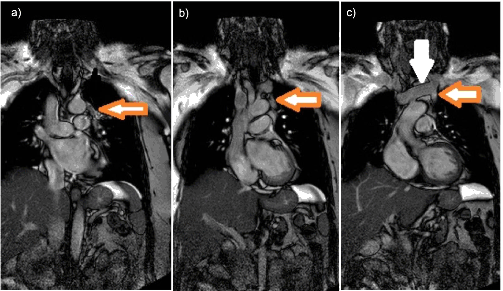
Legend: An MRA Scan Showing (a) An orange arrow points towards the left upper pulmonary vein
at the beginning of the anastomosis (b) The orange arrow highlights the middle of
the anastomosis between the left upper pulmonary vein and the brachiocephalic vein
(c) The arrow is pointing out the left upper pulmonary vein anastomosis with the brachiocephalic
vein, marked by the white arrow).
Six months after discharge, the patient was seen in the cardiology clinic and was
asymptomatic from the PAPVC, denying any shortness of breath or further chest pain.
Studies have shown asymptomatic PAPVC patients with low post-surgical repair mortality.13 One study showed that 93% of patients were free from arrhythmia medications at post-surgical
follow-up and the remaining 7% had either atrial fibrillation, sinus node dysfunction
(SND) or SND ectopic atrial rhythm. 13 Most common surgical risks included getting an arrhythmia, risks of hemorrhage, thrombus,
infection, poor tolerance of analgesic methods used and death. However, as these risks
are smaller than the risks of untreated PAPVC. Symptomatic patients or those with
complications of pulmonary hypertension and right ventricle hypertrophy should be
considered for surgical correction to prevent developing a LTRS and eventually RS
heart failure.12-14
Articles have previously reported diagnosing incidental PAPVC in patients with lung
cancer. One patient's PAPVC anatomy mimicked our patient and although asymptomatic,
they had increased mean arterial pressure and Qp/Qs ratio > 2.0. 15 Despite a significant co-morbidity of lung cancer, the post-surgical recovery and
survival was commendable and supportive of surgery. Despite follow up by cardiologists,
some patients were not diagnosed with PAPVC until post CT.16
These studies advocate using multimodality imaging to aid diagnosis of PAPVC especially
in symptomatic patients with chest symptoms. Ultimately, PAPVC is rare and further
research is required showcasing different presentations and ethnic groups.15-16
This case highlights incidental PAPVC found on a cardiac MRA performed for assessment
of a Ml. Our case was discussed in MDT and being asymptomatic from the PAPVC perspective,
the decision was not for surgery and to follow the patient in the cardiology outpatient
clinic. Symptomatic PAPVC patients can be managed by surgery and repair of the anomalous
connection may be performed successfully.
In conclusion, PAPVC is rare, can be fatal and in this case could be easily overlooked
as RS heart symptoms were absent. Awareness is crucial as if PAPVC patients become
symptomatic, surgical correction may be considered; hence appropriate follow up should
be accomplished. Ultimately this case emphasizes the importance of i) advocating relevant
multimodality imaging, ii) appropriate patient follow-up iii) and surgical consideration
for PAPVC patients who are symptomatic or have developed pulmonary or RS heart complications.
Key Points:
- PAPVC is a rare, often silent and can be easily missed which clinicians should be
aware of.
- Patients should be followed up closely for development of symptoms or worsening of
the LTRS.
- It is very important to systematically review all the data obtained from every imaging
modality to prevent missing a potential diagnosis.
Acknowledgments
None.
Conflict of Interest Statement & Funding
The Authors have no funding, financial relationships or conflicts of interest to disclose.
Author Contributions
Conception and design the work/idea: MW. Collect data/obtaining results MW. Analysis
and interpretation of data: MW. Write the manuscript: IQ, MW. Administrative or technical
advice: IQ. Critical revision of the manuscript: IQ, MW. Approval of the final version:
IQ, MW.
References
1.Haramati LB, Moche IE, Rivera VT, Patel PV, Heyneman L, McAdams HP, . Computed tomography of partial anomalous pulmonary venous connection in adults. J Comput Assist Tomogr. 2003 Sep-Oct;27(5):743–9.
2.Kottayil BP, Dharan BS, Menon S, Bijulal S, Neema PK, Gopalakrishnan SK, . Anomalous pulmonary venous connection to superior vena cava: Warden technique. Eur J Cardiothorac Surg. 2011 Mar;39(3):388–91.
3.Nakahira A, Yagihara T, Kagisaki K, Hagino I, Ishizaka T, Koh M, . Partial anomalous pulmonary venous connection to the superior vena cava. Ann Thorac Surg. 2006 Sep;82(3):978–82.
4.Kobayashi D, Williams DA, Cook AL. Mixed-type total anomalous pulmonary venous connection. Pediatr Cardiol. 2010 Aug;31(6):929–30.
5.Ho VB, Bakalov VK, Cooley M, Van PL, Hood MN, Burklow TR, . Major vascular anomalies in Turner syndrome: prevalence and magnetic resonance angiographic
features. Circulation. 2004 Sep 21;110(12):1694–700.
6.van den Hoven AT, Chelu RC, Duijnhouwer AL, Demulier L, Devos D, Nieman K, . Partial anomalous pulmonary venous return in Turner syndrome. Eur J Radiol. 2017 Oct;95:141–146.
7.Kadam S. Partial Anomalous Pulmonary Venous Connection. International Education and Research Journal. 2017 Sep; 3(9):20–21.
8.Babb JD, McClynn TJ, Pierce WS, Kirkman PM. Isolated partial anomalous venous connection: a congenital defect with late and serious
complications. Ann Thorac Surg. 1981 Jun;31(6):540
9.Sears EH, Aliotta JM, Klinger JR. Partial anomalous pulmonary venous return presenting with adult-onset pulmonary hypertension. Pulm Circ. 2012 Apr-Jun;2(2):250–5.
10.Edwin F. Left-sided partial anomalous pulmonary venous connection-should diagnosis lead to
surgery? Interact Cardiovasc Thorac Surg. 2010 Dec;11(6):847–8.
11.Uçar T, Fitoz S, Tutar E, Atalay S, Uysalel A. Diagnostic tools in the preoperative evaluation of children with anomalous pulmonary
venous connections. Int J Cardiovasc Imaging. 2008 Feb;24(2):229–35.
12.Prasad SK, Soukias N, Hornung T, Khan M, Pennell DJ, Gatzoulis MA, . Role of magnetic resonance angiography in the diagnosis of major aortopulmonary collateral
arteries and partial anomalous pulmonary venous drainage. Circulation. 2004 Jan 20;109(2):207–14.
13.Sahay S, Krasuski RA, Tonelli AR. Partial anomalous pulmonary venous connection and pulmonary arterial hypertension. Respirology. 2012 Aug;17(6):957–63.
14.Pace Napoleone C, Mariucci E, Angeli E, Oppido G, Gargiulo GD. Sinus node dysfunction after partial anomalous pulmonary venous connection repair. J Thorac Cardiovasc Surg. 2014 May;147(5):1594–8.
15.Asakura K, Izumi Y, Kohno M, Watanabe M, Arai T, Nomori H. Partial anomalous pulmonary venous connection associated with lung cancer in the same
lobe: report of a case. Ann Thorac Cardiovasc Surg. 2014;20 Suppl:457–60.
16.Takei H, Suzuki K, Asamura H, Kondo H, Tsuchiya R. Successful pulmonary resection of lung cancer in a patient with partial anomalous
pulmonary venous connection: report of a case. Surg Today. 2002;32(10):899–901.
Iqra Qamar, 1 University of Leeds, Leeds, United Kingdom
Mohammad Waleed, 2 Dr., Leeds General Infirmary, Leeds, United Kingdom
About the Author: Iqra Qamar is affiliated with the University of Leeds, Leeds, United Kingdom.
Correspondence: Mohammad Waleed, Address: Great George St, Leeds LSI 3EX, UK. Email: waleed1280@yahoo.com
Cite as: Qamar I, Waleed M. A Case Report Looking at an Incidental Finding of a Partial Anomalous Pulmonary Venous Connection Using Magnetic Resonance Angiography Int J Med Students. 2018 Jan-Apr;6(1):28-30.
Copyright © 2018 Iqra Qamar, Mohammad Waleed
International Journal of Medical Students, VOLUME 6, NUMBER 1, April 2018