Case Report
8-Year-Old Child with Cerebral Palsy Treated with Pelvic Osteotomies Using 3.5 mm
Blade Plate Having Subsequent Bilateral Implant Aseptic Loosening: A Case Report
Ahmed Nahian1, Julieanne P. Sees2
doi: http://dx.doi.org/10.5195/ijms.2021.698
Volume 9, Number 1: 52-55
Received 21 08 2020:
Rev-request 21 10 2020:
Rev-request 11 01 2021:
Rev-request 09 02 2021:
Rev-recd 19 11 2020:
Rev-recd 13 01 2021:
Rev-recd 10 02 2021:
Accepted 09 04 2021
ABSTRACT
Background:
Cerebral palsy (CP) is a central problem of the brain due to neurological insult that
affects muscle posture, tone, and movement, resulting in poor motor control and dysfunctional
muscle balance affecting hip joints in the growing child. Surgical treatment of hip
and, if present, acetabular dysplasia addresses the femoral neck-shaft angle, appropriate
muscle lengthening, and deficiency of acetabular coverage, as necessary. The surgeons
perform proximal femoral osteotomies (PFOs) mostly with fixed angled blade plates
(ABP) with proven success. The technique using an ABP is common and requires detailed
attention to perform and to teach.
The Case:
In this case, an eight-year-old ambulatory patient with CP underwent bilateral proximal
varus femoral derotational and pelvic osteotomies for the neuromuscular hip condition
with a 3.5 mm Locking Cannulated Blade System (OP-LCP) by OrthoPediatrics Corp instead
of the use of the conventional 4.5 mm ABP procedure, resulting in aseptic loosening.
Conclusion:
Due to the child's underdeveloped posture, the surgeon utilized the 3.5 mm instrumentation
for a child-size implant, which worked sufficiently for the surgery but may not have
loosened if a similar child-size blade plate system of 4.5 mm screws was implanted.
While the ABP and OP-LCP systems are effective and safe for internal corrections of
PFOs, the OP-LCP system may aid the residents in learning the procedure with higher
confidence, fewer technical inaccuracies, and refined outcomes. Both systems are safer
and viable for the treatment of neuromuscular hip conditions.
Keywords:
Cerebral Palsy;
Hip Dislocation;
Osteotomy;
Gait;
Acetabuloplasty;
Bone Anteversion (Source: MeSH-NLM).
Highlights:
- Cerebral palsy (CP) is the most prevalent motor disability in childhood.
- Hip dysplasia, caused by CP, can be avoided by early screening, and fixed at a lower
risk with procedural measures, i.e., osteotomies, on children with alarming hips.
- Though fruitful, the angled blade plate method of performing osteotomies is unnecessarily
complex, while modern technology allows for more viable methods.
Introduction
Cerebral palsy (CP) is a central problem of the brain due to neurological insult that
affects muscle posture, tone, and movement, causing poor motor control and affecting
the extremities, particularly the forces of the hip joint. In a review of children
with the hip disease across all Centers for Disease Control and Prevention (CDC) sites,
researchers found that 3.1 prevalence per 1,000 children were born with CP.1 Children with CP are prone to develop hip dysplasia. Often confused with congenital
dysplasia of the hip (CDH), children with CP-led hip dysplasia have different development
pathways. Clinical diagnosis of CDH is carried with findings of a lax dislocated hip
at birth, tighter displaying hip at six weeks postpartum, or an abnormal gait at 15
weeks postpartum. Common symptoms of CDH include asymmetrical or abducted legs along
with a limited range of motion in squatting, walking, and crawling positions.2,3 In patients with CP, the hip is usually normal at birth, but the hampered motor development
leads to dysplasia. Gross Motor Function Classification System (GMFCS) tracks movements
like walking, sitting, and mobile device usage to deliver a lucid description of a
child's updated motor function and a concept of what mobility aid a child may require,
e.g., walking frames, crutches, and wheelchairs. The rate of hip displacement in children
with CP has been demonstrated as a linear relationship with the child's GMFCS level.4
Surgical correction of hip dysplasia commonly involves addressing the femoral neck-shaft
angle, appropriate muscle lengthening, and addressing acetabular coverage deficiency
as necessary.5,6 Proximal femoral osteotomies (PFOs) are mostly performed with fixed angled blade
plates (ABP) with proven success, while instrumentation of proximal femoral locking
plate systems is often sufficient. The osteotomy technique using an ABP, however,
is complex to perform and to teach.6 In this case, we present an eight-year-old patient with CP who underwent bilateral
hip varus femoral derotational and pelvic osteotomies for hip dysplasia with a 3.5
mm Locking Cannulated Blade System (OP-LCP) by OrthoPediatrics Corp. The patient's
smaller size dictated usage of the 3.5 mm LCP for that manufacturer, which would be
comparable to more conventional 4.5 mm ABP such as other systems manufactured; thus,
the nature of a less robust implant strength likely resulted in the aseptic loosening.
The Case
An eight-year-old ambulatory male patient GMFC III was found to have an increasing
hip subluxation and acetabular dysplasia despite continued progress with a posterior
walker and a previous history at age five of bilateral hip adductor, iliopsoas, and
gracilis muscles lengthening. In general, a pediatric patient of his age and GMFCS
level has 27% success of not developing further hip subluxation after muscle lengthenings.7 Upon radiographic (Figure 1) review, as confirmed on computed tomography scan, bilateral uncovering of the femoral
heads with right acetabular dysplasia posteriorly was observed. As the patient was
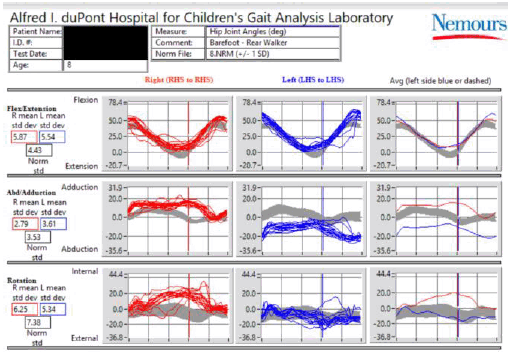
ambulatory, a 3D instrumented gait analysis was conducted demonstrating bilateral
mild hip internal rotation, normal flexion/extension motion, and mild lurching secondary
to bilateral abductor weakness and poor control (Figure 2).
Figure 1.
Preoperative X-Ray Showing CP-Caused Hip Dysplasia in Supine Position. Anterior to
Posterior (Front to Back) X-Ray Displaying Hip Dysplasia in the Right Hip (at the
Orange Arrow). The Dotted Red Lines Show the Shortage of Acetabular Coverage in the
Dysplastic Hip vs. the Healthy Hip.
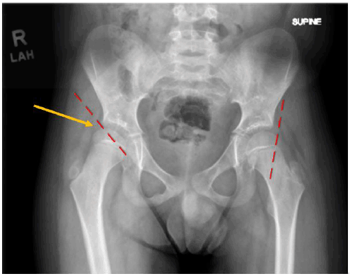
Figure 2.
3D Instrumented Gait Analysis of Hip Motion During Ambulation for an 8-Year-Old Boy
GMFC III with CP. The Patient is Seen with Out-of-Range Motion Compared Against Normal
Standard Deviations.
With evidence of physical exam, radiographs, and 3D gait analysis, this child was
sent to a pediatric neuromuscular-orthopedic surgeon, who performed bilateral hip
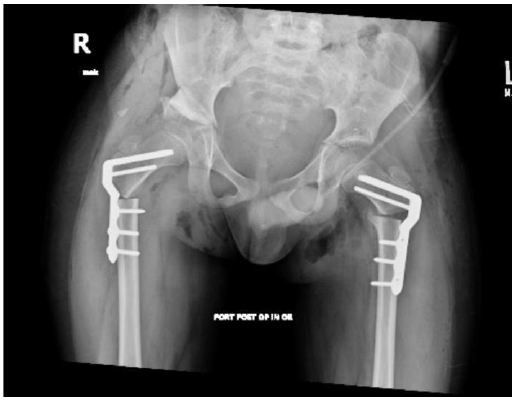
femoral varus derotational osteotomies with right acetabuloplasty (Figure 3). Imaging and clinical findings dictated preoperative planning for PFOs. The patient
was in the supine position during surgery; the patient's position and closure are
the same in most PFOs. The 5 cm long incision was laterally originating at the tip
of the greater trochanter. The femoral osteotomy goal was to provide a medially based
closing wedge osteotomy. The aim was to improve the hip's centering by restoring the
femoral neck and head by decreasing (varus) the femoral neck-shaft angle. Internal
fixation commonly requires an ABP or the LCP (Locking Cannulated Blade System). LCP
supplies primary strength by locking screws on a plate and deviation of femoral neck
screws and involves guidewires. The guidewire for the blade plate chisel was inserted
into the femoral neck along its axis. Since the varus correction was conducted at
30° with a 110° plate, the guidewire was placed by adjusting the instruments to 140°.
After placing the child-size chisel, due to fluoroscopy, particularly in the lateral
intraoperative view, the lateral femur was too small and unable to accommodate the
adolescent 4.5 mm cannulated system. The medially based femoral wedge osteotomy was
performed; the OP plate was then placed into the proximal fragment without difficulty,
and the osteotomy was reduced, assuring bilateral symmetry of length and rotation,
affixed to the femur. Final intraoperative radiography was satisfactory upon completing
all procedures, including pelvic osteotomy of the right hip for adequate bilateral
hip reconstruction.
Figure 3.
Immediate Postoperative X-Ray After Initial Hip Surgery. The Bones Have Been Spliced
and Conjoined with LCP.
The patient tolerated the procedure and was admitted for a 6-week course of inpatient
rehabilitation. His full weight-bearing status was tolerated immediately in postoperative
conditions; after one year of the surgery, osteotomies were healed, and no complaints
were reported with his ambulatory functional level maintained (Figure 4). Due to his young age at the time of implantation, the initial plan was to remove
the implants at one year postoperative. At the one-year postoperative clinic visit,
the parents were unclear if there were any painful symptoms from the hardware prominence
due to the child's limited communicative ability. According to the patient's mother,
in retrospect, he had a vague thigh pain as he would point anteriorly or laterally
to his thighs intermittently after more extended periods of walking and upright activities.
The family proceeded with outpatient surgical hardware removal at 18-months postoperative
with intraoperative findings marked with significant trochanteric bursitis bilaterally
and symmetric bilateral loosening of shaft screws (Figure 5). The child subsequently tolerated the hardware removal without any complications,
returning to his previous functional level, pain-free.
Figure 4.
Radiograph at 18-Months in Postoperative, the Patient Displays Healed Osteotomies;
The Distal Femoral Shaft Screws Are Backing Out Bilaterally.
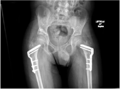
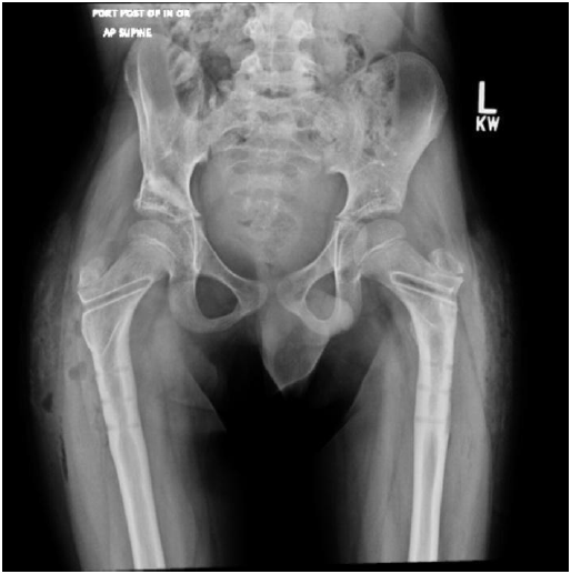
Figure 5.
Postoperative X-Ray After Implant Removal.
Discussion
The treatment of hip contractures caused by cerebral palsy was first published in
1880.8 A retrospective review indicates a lateral dislocation (partial) and subluxation
of the hip existing in 30-60% of non-ambulatory ambulatory pediatric CP patients aged
five years.9 The patient presented in this case was of GMFCS level III, thus using an assistive
device for his ambulation. Being an ambulatory child with a gait abnormality, notable
lurching, and a combination of poor motor control and along with movements of the
iliotibial band abductor complex, the smaller nature of the 3.5 mm system over a 4.5
mm stronger plate setup may have led to mechanical implant loosening after femoral
osteotomies healed.
The standard protocol is to determine the level of dislocation or subluxation of the
hip joint using an anteroposterior radiograph, as used in this case, to measure hip
migration percentage. While this case may not have involved many patients, the surgical
and radiographic procedures were standard. The pelvic and hip positions during radiography
were consistent with verifying the reliable change. In previous cases, the migration
rate often indicated the level of risk—a 7% annual migration corresponded with a developing
hip disability. A displacement between 33% and 80% indicates hip subluxation, and
over 80% indicates hip dislocation. The interconnection between hip migration and
migration percentage is relatively vague, and migration percentage is not uniformly
indicative of a child's clinical and functional picture, which eventually plays a
significant role in treatment plans. A bilateral check on hip integrity after an intervention
is suggested; bilateral functionality and pain-free range of motion (ROM) have always
acted as the benchmark for surgical success. In ambulatory children with notable symptoms
of internally rotated gait secondary to internal rotation at the hip, the prodigal
time for reconstruction is between 5 and 7 years.10 The risk of establishing recurrent bony anteversion is much lower around that age
range; furthermore, motor control, planning, and balance start to reach their peak
at this range. This peak allows for full restoration of function by formal elementary
education, crucial to middle childhood.10
While CP and hip deformity are commonly treated conditions, the surgeon utilized a
relatively newer but proven effective surgical hip fixation method. Dating back to
the time of Hippocrates, osteotomies are generally performed for two reasons: to precisely
realign a bone's axis or to allow bone transport.11 As used in this case, a simple osteotomy may fix rotational or angular defects by
which healing is in compression. This procedure solely depends on stability to encourage
union in the postoperative position. As mentioned earlier, PFOs are mostly performed
with fixed angled blade plates (ABP) due to proven success rate and lower cost. Despite
its positive results, ABP is technically challenging for those accustomed to the implant
and requires experienced attention. In this patient's case, the surgeon utilized the
3.5 mm instrumentation for a child-size implant, which worked sufficiently for the
surgery but may not have loosened if a similar child-size blade plate system of 4.5
mm screws was implanted. OP-LCPs are relatively more expensive than ABPs; however,
LCPs are better for steadiness, angle correction, and in training institutions in
treatment of neurological hip disorders, such as CP.12 Based on the results of this case, we conclude that both the ABP and OP-LCP systems
are fruitful and safe for internal corrections of PFOs (Table 1). Additionally, the OP-LCP system may aid the residents in learning the procedure
with higher confidence, fewer technical inaccuracies, and refined outcomes; this case
brings caution to using this system in all hip deformities of ambulatory children.
OP-LCP is exclusively targeted for the surgical correction of pediatric hip deformity,
fixed knee flexion deformity, and trauma. The system acts as a primary exposure to
osteotomies for trainees via easy-to-use locking screws in the proximal and distal
fragments. OP-LCP employs various offsets to restore the mechanical axis of the lower
limb, allowing the surgeons to use unique methodology but produce reproducible results.
Finally, the implant removal difficulty level must be cautiously reviewed when selecting
a method of performing pediatric PFOs. Removal of all blade plates requires attention,
knowing there is a high prevalence of fractures and retained hardware in children
with CP.13 The removal of the OP-LCP, in this case, was safely performed and extracted without
morbidity, which is supported by previous literature.
Table 1.
Procedural Comparison Between ABP and OP-LCP Methods.
| Step |
ABP |
OP-LCP |
| Guidewire insertion |
approximated to line of the seating chisel |
precise to line of the seating chisel |
| Chisel insertion |
below guidewire, which serves only as a reference plane |
directly over the wire |
| Implant insertion |
approximate and adjustable |
plannable, predictable, and precise |
Acknowledgments
The authors acknowledge Nemours/Alfred I. duPont Hospital for Children, United States.
Conflict of Interest Statement & Funding
The Authors have no funding, financial relationships or conflicts of interest to disclose.
Author Contributions
Conceptualization, Methodology, Project Administration, Visualization, Writing – Original
Draft, & Writing – Review and Editing: AH & JPS. Investigation, Resources, & Supervision:
JPS.
References
1.
Centers for Disease Control and Prevention. Data and Statistics for Cerebral Palsy. Available from: https://www.cdc.gov/ncbddd/cp/data.html. Last updated Apr 30, 2019; cited Aug 4, 2020.
2.
International Hip Dysplasia Institute. Developmental Dysplasia of the Hip (DDH). Available from: https://hipdysplasia.org/developmental-dysplasia-of-the-hip/. Last updated Jul 4, 2020; cited Aug 4, 2020.
3.
International Hip Dysplasia Institute. FAQ #4 What Are Some Signs I Can Look for if I Suspect I Have Hip Dysplasia? Available from: https://hipdysplasia.org/developmental-dysplasia-of-the-hip/faq/#:~:text=Hip%20dysplasia%20that%20needs%20treatment,common%20in%20girls%20than%20boys. Last updated Jul 4, 2020; cited Aug 4, 2020.
4. Soo B, Howard JJ, Boyd RN, Reid SM, Lanigan A, Wolfe R, et al. Hip displacement in cerebral palsy. J Bone Joint Surg Am. 2006 Jan;88(1):121–9.
5. Dyson PH, Lynskey TG, Catterall A. Congenital hip dysplasia: problems in the diagnosis and management in the first year
of life. J Pediatr Orthop. 1987 Oct;7(5):568–74.
6. Miller, F. Cerebral palsy. 1st ed. New York: Springer–Verlag; 2005.
7. L. Zhou, M. Camp, A. Gahukamble, K. L. Willoughby, M. Harambasic, C. Molesworth, et al. Proximal femoral osteotomy in children with cerebral palsy: the perspective of the
trainee. J Child Orthop. 2017 Jan;11(1):6–14.
8. Reimers J. The stability of the hip in children. A radiological study of the results of muscle
surgery in cerebral palsy. Acta Orthop Scand Suppl. 1980 Dec;51Suppl184:S1–100.
9. Shore B, Spence D, Graham H. The role for hip surveillance in children with cerebral palsy. Curr Rev Musculoskelet Med. 2012 Jun;5(2):126–34.
10. Kazemi SM, Minaei R, Safdari F, Keipourfard A, Forghani R, Mirzapourshafiei A. Supracondylar Osteotomy in Valgus Knee: Angle Blade Plate Versus Locking Compression
Plate. Arch Bone Jt Surg. 2016 Jan;4(1):29–34.
11. Dabis J, Templeton-Ward O, Lacey AE, Narayan B, & Trompeter A (2017). The history, evolution and basic science of osteotomy techniques. Strategies in trauma and limb reconstruction. 2017 Oct 06;12(3):169–80.
12. Khouri N, Khalife R, Desailly E, Thevenin-Lemoine C, Damsin JP. Proximal femoral osteotomy in neurologic pediatric hips using the Locking Compression
Plate. J Pediatr Orthop. 2010 Dec;30(8):825–31.
13. Pountney T, Green EM. Hip dislocation in cerebral palsy. BMJ. 2006 Mar 30;332(7544):772–5.
Ahmed Nahian, 1 BS/DO (c), California Baptist University - Lake Erie College of Osteopathic Medicine,
United States
Julieanne P. Sees, 2 FAAOS, FAOA, FAOAO, AI DuPont Hospital for Children, United States
About the Author: Ahmed Nahian is currently a second-year BS/DO student at California Baptist University
– Lake Erie College of Osteopathic Medicine, Erie, USA of an eight-year medical school
program. He is also a recipient of the prestigious Parker B. Francis Fellowship in
Pulmonary Critical Care Research administered by the University of Kansas Medical
Center (KUMC) and is deeply interested in the musculoskeletal system and prosthetic
science, as he wants to pursue orthopedic surgery after medical school.
Correspondence: Ahmed Nahian, Address: 8432 Magnolia Ave, Riverside, CA 92504, United States. Email:
ahmed.nahian.usa@gmail.com
Editor: Ryan T. Sless
Student Editors: Lourdes Adriana Medina-Gaona, Sohaib Haseeb & Azher Syed
Copyeditor: Leah Komer
Proofreader: Madeleine Jemima Cox
Layout Editor: Annora Ai-Wei Kumar
Cite as: Nahian A, Sees JP. 8-Year-Old Child with Cerebral Palsy Treated with Pelvic Osteotomies Using 3.5 mm Blade Plate Having Subsequent Bilateral Implant Aseptic Loosening: A Case Report. Int J Med Students. 2021 Jan-Apr;9(1):52-5.
Copyright © 2021 Ahmed Nahian, Julieanne P. Sees
This work is licensed under a Creative Commons Attribution 4.0 International License.
International Journal of Medical Students, VOLUME 9, NUMBER 1, April 2021


