Anatomy of the Cerebellum.
Claudia K. Sellers1, Suvankar Pal2
doi: http://dx.doi.org/10.5195/ijms.2013.29
Volume 1, Number 1: 37-42
Received 04 01 2012: Accepted 10 07 2012
ABSTRACT
The cerebellum is central to normal motor function and co-ordination, and can be frequently affected in a number of common disease processes. However, medical student teaching relating to cerebellar anatomy and pathology is lacking, leaving many graduates with a significant knowledge gap. Junior doctors need to be able to recognize ‘cerebellar syndromes’ on presentation to hospitals, and to identify and manage reversible causes rapidly and effectively. After review of relevant literature, a simple approach to the functional anatomy and practical classifications of common cerebellar pathology is presented here, with a focus on symptoms, signs and examination techniques essential to medical school final exams.
Keywords: Cerebellar Diseases; Central Nervous System Diseases; Cerebellum; Students, Medical.
The cerebellum (Latin for ‘little brain’) is located infero-posteriorly to the cerebral cortex, and is fundamental to normal neurological functioning, yet it was not until the early 20th Century that Flourens discovered its primary function to be in motor control and co-ordination.1 The cerebellum may be affected in common neurological disorders such as stroke, multiple sclerosis and mass lesions, often producing a ‘cerebellar syndrome’. Junior doctors need to be confident in recognition and investigation of patients presenting with cerebellar symptoms and signs appropriately in order to identify treatable causes and provide accurate prognosis. Textbook coverage of this area is notoriously disjointed, often leaving medical students with a significant knowledge gap. This article aims to provide a brief overview of the etiology of cerebellar disorders and a simple approach to history, examination and investigation of patients presenting with cerebellar syndromes.
A review of literature was performed using PubMed, Google Scholar, Embase and Medline databases for articles from 1975 to 2012 using the search terms: Cerebellum, Neurological Examination, Ataxia, and Cerebellar Syndromes. Additional papers were retrieved from reviews and references. Articles included were in the English language and related to anatomy and clinical examination technique of the cerebellar system.
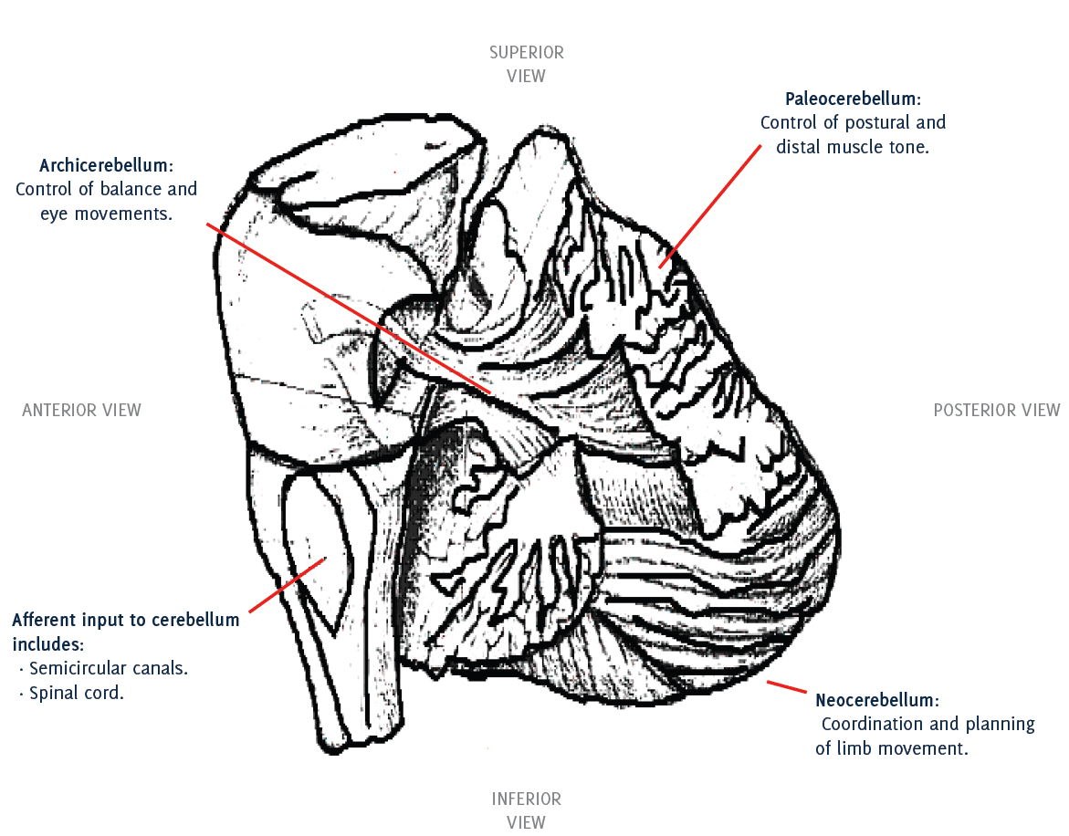
The cerebellum is attached to the dorsal aspect of the brainstem by three separate fibrous structures - the superior, middle and inferior peduncles, which connect to the mid-brain, pons and medulla respectively (Figure 1). It comprises two hemispheres joined in the midline by the ‘vermis’, which, unlike the cerebral hemispheres, are ipsilateral in their control of co-ordination. Afferent input to the cerebellum includes neuronal tracts from the semicircular canals, proprioception via the spinal cord, motor information from the frontal lobes, and fibers from reticular nuclei in the brainstem. Efferent output is to midbrain and thalamic nuclei, or directly to the motor cortex, permitting feedback comparing sensory and motor input with output.
Figure 1.Anatomy of the Cerebellum.
The anatomy of the cerebellum can also be further divided functionally. Control of balance, posture and eye movements is mediated by the flocculonodular lobe (or archicerebellum) which connects with the vestibular and reticular nuclei in the brainstem. The vermis and paravermis (the intermediate section between the cerebellar hemispheres) together make up the paleocerebellum, responsible for control of postural and distal muscle tone. Finally, coordination and planning of limb movement occurs within the neocerebellum, which is comprised of the remaining cerebellar hemispheres.2
Cerebellar pathology arises secondary to a broad range of etiologies which can be usefully classified according to the time-course of symptoms:
Usually cerebellar infarction or hemorrhage. This may be associated with headache, vertigo, vomiting and altered consciousness.
Usually transient and last minutes to hours, there are various causes:
A ‘surgical sieve’ approach to the commoner causes is illustrated in Table 1.
Table 1.‘Surgical Sieve’ approach to common causes of cerebellar syndromes.
| Vascular | Stroke (infarct or hemorrhage)*, Transient ischemic attack* |
| Inflammatory | Multiple sclerosis* |
| Neoplastic | Primary tumors: Astrocytoma, Medulloblastoma, Haemangioblastoma |
| Secondary tumors: metastases* (commonly lung, breast and GI tract) | |
| Paraneoplastic phenomena (Anti Hu Ab in small cell lung cancer) (rare) | |
| Toxic/Trauma | Alcohol* |
| Metabolic | Hypoglycemia*, Hypoxia*, Hypothyroidism*, thiamine deficiency* |
| Infectious | Bacterial: Meningo-encephalitis, Intracranial abscess, Viral: Varicella, HIV |
| Parasitic infections (rare): Toxoplasma, Falciparum Malaria | |
| Congenital | Agenesis, Dandy-Walker malformation, Arnold-Chiari malformation (rare) |
| Inherited | Friedrich’s Ataxia, spinocerebellar ataxias Degenerative Multisystem atrophy, Spinocerebellar ataxias, prion disease (rare) |
| Drugs | Barbiturates*, Phenytoin and other Anticonvulsants*, Anti-neoplastic drugs*. |
The cerebellum operates at a subconscious level to control muscle tone, posture and co-ordination. Cerebellar disorders result in difficulties with the rate, rhythm and force of limb movements, gait and speech.3 The key symptoms and signs can be remembered using the mnemonic DANISH:
Patients may also present with symptoms relating to underlying pathology for example, headache, nausea and vomiting resulting from a cerebellar tumor; systemic upset associated with a cerebellar abscess, and pyramidal and sensory signs in multiple sclerosis.
A guide to comprehensive examination of cerebellar function as may be expected in an undergraduate Objective Structured Clinical Examination (OSCE) is presented in Table 2.
Table 2.A systematic approach to cerebellar examination. ‘Please examine this patient’s cerebellar function’. How to pass the OSCE.
| Action | Potential Findings | |
|---|---|---|
| Introduction | Wash your hands. Introduce, explain, consent the patient. Position the patient at 45° lying down. |
|
| Inspection | Bruising, scars ‘bobbing ‘ head Walking aids | Recurrent falls Titubation gait ataxia |
| Head | ||
| Nystagmus | Follow your finger with eyes, head still | Broken pursuit, nystagmus the degree of involuntary eye movement is greatest when gaze is focused to the same side as the lesion. |
| Speech | Ask to repeat ‘West Register Street, British Constitution, PP PP PP, KK KK KK, TT
TT TT’ Ask to read a sentence aloud |
Staccato Speech Slurring |
| Limbs | ||
| Tone | As in normal neurological exam | May be increased after a stroke or in multiple sclerosis. |
| Power | As in normal neurological exam | May be reduced with associated upper motor neuron pathology. ↓ power may confound incoordination |
| Coordination | UPPER LIMBS Test for ‘rebound’: -Arms out straight in front with eyes closed, push each arm down ~ 10cm then release6 |
Overshooting = Dysmetria |
| Finger-nose Test Examiner holds index finger about 50cm in front of the patient’s face, and asks the patient to go between touching his/her nose and the examiner’s finger as accurately as possible. |
Difficulty = Dysmetria Intention tremor, past pointing | |
| Hand-turning test Ask the patient to tap one hand on the back of the other, and then tap the same spot with the dorsum of the same hand. |
Difficulty = Dysdiadochokinesis | |
| Coordination | LOWER LIMBS Heel-shin test Put heel onto opposite knee and slide heel down shin to ankle. Lift off and repeat. |
Intention tremor Difficulty = Dysmetria |
| Foot tapping Ask to tap foot as rapidly as possible |
Difficulty = Dysdiadochokinesis | |
| Posture and Gait | ||
| Posture | Ask how stable they are sitting/standing? Assess sitting down Also with arms crossed |
Truncal ataxia |
| Assess standing (If stable sitting) Arms by sides, feet together |
Truncal ataxia | |
| Gait | Ask to walk across room Ask to walk heel to toe |
Wide-based gait (midline cerebellar lesions) Unsteadiness and lateral veering (hemispheric lesions) Irregular steps |
| Concluding Remarks | “I would also like to carry out a full neurological examination” Investigations: MRI posterior fossa, and specific as indicated (see text) |
Handy Hints:
“Overall, specific tests can be helpful in confirming or refuting the presence of cerebellar disease, but must be interpreted in the context of other signs and the case history.”
Investigation should be guided according to the differential diagnoses in mind, but commonly includes blood tests and cerebellar imaging.
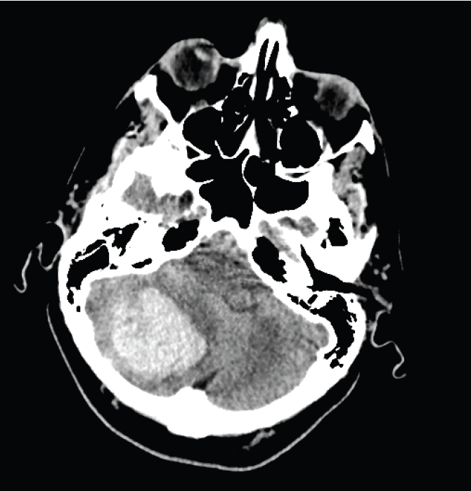
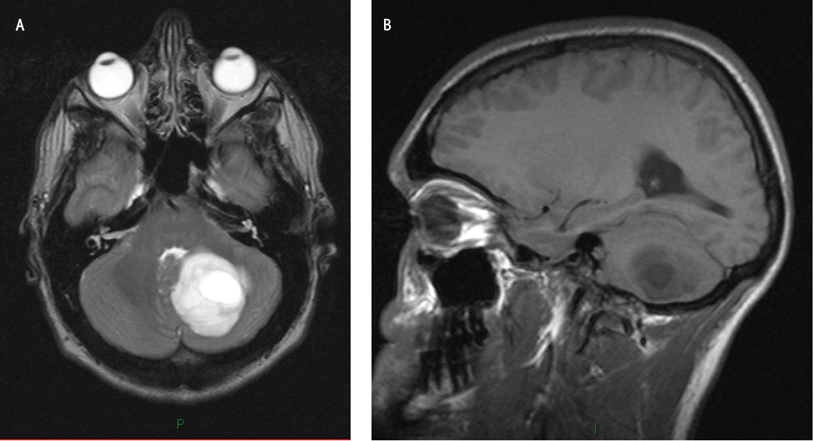
Imaging: Computed tomography (CT) (Figure 2) or Magnetic resonance tomography (MRI) (Figure 3) of the brain are used to investigate cerebellar disorders and may reveal a stroke, space occupying lesion, demyelination or atrophy.
Figure 2.CT head scan showing right sided cerebellar hemorrhage. This elderly patient presented with sudden onset headache, nausea, vomiting, unsteady gait and right sided clumsiness.
A (axial section) and B (sagittal section). MRI head scan showing mixed solid/cystic left cerebellar hemisphere mass with hydrocephalus and cerebellar tonsillar herniation caused by a pylocytic astrocytoma. This young patient presented with a subacute left sided cerebellar syndrome associated with headache.
The cerebellum is affected by a number of common disease processes, producing characteristic symptoms and signs. Junior doctors need to be able to accurately detect such ‘cerebellar syndromes’ since they are a common presentation to hospital and since there are a number of treatable causes which can be reversed if detected early.5 A sound understanding of the anatomy of the cerebellum, etiology of cerebellar disorders and logical approach to examination will facilitate appropriate investigation and treatment.6
Investigations: Blood tests, Imaging, CSF examination and genetic screening as appropriate.
CJD, Creutzfeld-Jacob Disease
CSF, Cerebrospinal Fluid
CT, Computed Tomography
CXR, Chest X-ray
GI, Gastrointestinal
HIV, Human Immunodeficiency Virus
MRI, Magnetic Resonance Imagin
OSCE, Objective Structured Clinical Examination
TIA, Transient Ischemic Attack
The Authors have no funding, financial relationships or conflicts of interest to disclose.
1. Fine EJ, Ionita CC, Lohr L. The history of the development of the cerebellar examination. Semin Neurol 2002; 22(4):375–84
2. Levy MN, Koeppen BM, Stanton BA. Special Senses: The cerebellum assists in the regulation of posture and movement. In: Levy MN, Koeppen BM, Stanton BA (4th ed). Principles of Physiology. Philadelphia: Elsevier Mosby; 2006.
3. Shardlow A, Turner MR. Examination of the Cerebellum. J Clin Exam 2008; 5:5–9.
4. Khasnis A, Gokula RM. Romberg’s test. J Postgrad Med 2003; 49(2):169–72.
5. Manto M. The cerebellum, cerebellar disease and cerebellar research-two centuries of discoveries. Cerebellum 2008;7(4):505–16.
6. Angel RW. The rebound phenomenon of Gordon Holmes. Arch Neurol 1977; 34(4):250.
Claudia K. Sellers, 1 Department of Acute General Medicine, St Thomas’ Hospital, London, UK.
Suvankar Pal, 2 Department of Clinical Neurosciences, Western General Hospital, Edinburgh, Scotland.
About the author. Claudia Sellers is a Foundation Year 1 Doctor at Guy’s and St Thomas’ NHS Trust, London, UK, and graduated from the University of Edinburgh Medical School in 2011.
Correspondence Dr. Claudia Sellers. Address: Department of Acute General Medicine, St Thomas’ Hospital, London SE1 7EH. E. mail: claudiesellers@hotmail.co.uk
Cite as: Sellers CK, Pal S. Cerebellar Syndromes: A Medical Student Guide. Int J Med Students 2013;1(1):37-42.
Copyright © 2013 Claudia K. Sellers, Suvankar Pal
International Journal of Medical Students, VOLUME 1, NUMBER 1, April 2013