Cholecystocolonic Fistula: Demonstrating the Need for Further Imaging Assessment Following an Abnormal Ultrasound Exam
DOI:
https://doi.org/10.5195/ijms.2022.1831Keywords:
Ultrasound, Diagnostic, Intestinal Fistula, Colonic Neoplasms, Adenocarcinoma, Incidental FindingsAbstract
Background
Point of care ultrasound (PoCUS) is a diagnostic tool that can efficiently answer targeted clinical questions at the bedside. Such questions include confirming or ruling out the presence of a specific complication suspected by the clinician, like an abdominal aortic aneurysm, for example. Proper identification of any such complication is reliant upon a fundamental knowledge and recognition of normal anatomy in each view, so the ultrasound provider can distinguish normal from a variety of hallmark pathologic signs. A positive finding warrants immediate changes in management, often including further imaging to guide interventions. However, indeterminate, or incidental findings unrelated to the patient’s chief complaint can be found. While usually benign, sometimes these findings are indicative of an underlying pathology not initially suspected by the physician. In these settings, PoCUS has limited diagnostic value, and therefore it is important to highlight the need for further imaging following discovery of abnormal or incidental findings on an ultrasound exam.
Case
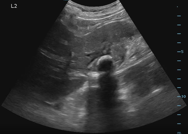
The patient was a 75-year-old female with COPD, coronary artery disease and hypertension. Her overall health declined after an admission for COVID pneumonia, which required treatment for oxygen. She never improved completely and was diagnosed with pulmonary fibrosis, likely secondary to COVID-19. She presented to our outpatient clinic for follow up from a recent hospitalization for respiratory decompensation and heart failure. During the visit she complained of intermittent right sided abdominal pain which had been present for a couple weeks. It was not associated with eating, and the pain did improve some after passing gas. The decision was made to perform a bedside ultrasound of her gallbladder to look for gallstones. Upon visualizing her gallbladder, hyperechoic shadowing in a smooth, circumferential nature filled the gallbladder. The differential included porceline gallbladder, stone filled gallbladder, or emphysematous cholecystitis. She was referred for further imaging, but before she could get imaging completed, she presented to the emergency department due to worsening pain. A CT scan of the abdomen showed an ill-defined soft tissue mass with surrounding inflammation involving the inferior right hepatic lobe, gallbladder and cecal visualization. Overall, given the surrounding inflammation this was favored to represent perforated cholecystitis with inflammatory fistula. Interventional radiology attempted to place a drain which was unsuccessful but did demonstrate fistulization with the colon. She later had a cholecystectomy performed, with a pathology report which detailed results showing metastatic poorly differentiated adenocarcinoma with signet ring and mucinous features. Oncology was consulted for treatment options, but unfortunately the patient passed away from cardiopulmonary compromise before treatment could be initiated.
Conclusion
This case demonstrates the importance of follow up imaging for abnormal bedside ultrasound studies which do not follow the typical PoCUS pathway. Point of care ultrasound is used to answer a binary question, “Does my patient have a gallstone?” for example. If there are abnormal findings, or findings which do not correlate with the history and physical examination, more advanced imaging assessment is required and should be ordered by the point of care ultrasound provider. 
Downloads
Published
How to Cite
Issue
Section
Categories
License
Copyright (c) 2022 Andrew J. Gauger, James Wilcox

This work is licensed under a Creative Commons Attribution 4.0 International License.
Authors who publish with this journal agree to the following terms:
- The Author retains copyright in the Work, where the term “Work” shall include all digital objects that may result in subsequent electronic publication or distribution.
- Upon acceptance of the Work, the author shall grant to the Publisher the right of first publication of the Work.
- The Author shall grant to the Publisher and its agents the nonexclusive perpetual right and license to publish, archive, and make accessible the Work in whole or in part in all forms of media now or hereafter known under a Creative Commons Attribution 4.0 International License or its equivalent, which, for the avoidance of doubt, allows others to copy, distribute, and transmit the Work under the following conditions:
- Attribution—other users must attribute the Work in the manner specified by the author as indicated on the journal Web site; with the understanding that the above condition can be waived with permission from the Author and that where the Work or any of its elements is in the public domain under applicable law, that status is in no way affected by the license.
- The Author is able to enter into separate, additional contractual arrangements for the nonexclusive distribution of the journal's published version of the Work (e.g., post it to an institutional repository or publish it in a book), as long as there is provided in the document an acknowledgment of its initial publication in this journal.
- Authors are permitted and encouraged to post online a prepublication manuscript (but not the Publisher’s final formatted PDF version of the Work) in institutional repositories or on their Websites prior to and during the submission process, as it can lead to productive exchanges, as well as earlier and greater citation of published work. Any such posting made before acceptance and publication of the Work shall be updated upon publication to include a reference to the Publisher-assigned DOI (Digital Object Identifier) and a link to the online abstract for the final published Work in the Journal.
- Upon Publisher’s request, the Author agrees to furnish promptly to Publisher, at the Author’s own expense, written evidence of the permissions, licenses, and consents for use of third-party material included within the Work, except as determined by Publisher to be covered by the principles of Fair Use.
- The Author represents and warrants that:
- the Work is the Author’s original work;
- the Author has not transferred, and will not transfer, exclusive rights in the Work to any third party;
- the Work is not pending review or under consideration by another publisher;
- the Work has not previously been published;
- the Work contains no misrepresentation or infringement of the Work or property of other authors or third parties; and
- the Work contains no libel, invasion of privacy, or other unlawful matter.
- The Author agrees to indemnify and hold Publisher harmless from the Author’s breach of the representations and warranties contained in Paragraph 6 above, as well as any claim or proceeding relating to Publisher’s use and publication of any content contained in the Work, including third-party content.
Enforcement of copyright
The IJMS takes the protection of copyright very seriously.
If the IJMS discovers that you have used its copyright materials in contravention of the license above, the IJMS may bring legal proceedings against you seeking reparation and an injunction to stop you using those materials. You could also be ordered to pay legal costs.
If you become aware of any use of the IJMS' copyright materials that contravenes or may contravene the license above, please report this by email to contact@ijms.org
Infringing material
If you become aware of any material on the website that you believe infringes your or any other person's copyright, please report this by email to contact@ijms.org


