Bronchiectasis with Transmediastinal Herniation of the Left Upper Lobe in a 3-Year-Old Child: A Case Report
DOI:
https://doi.org/10.5195/ijms.2024.2176Keywords:
Tuberculosis, Pulmonary medicine, Pediatrics, Bronchiectasis, Transmediastinal Herniation, High-Resolution Computed Tomography, Pediatric Respiratory Disease, Pectus Carinatum, Pulmonary Tuberculosis, Airway Inflammation, Antibiotic Therapy, Chest Radiograph, Respiratory DistressAbstract
Background: Bronchiectasis is a disorder marked by the destruction of smooth muscle and elastic tissue caused by inflammation, resulting in the permanent expansion of bronchi and bronchioles. It can occur following a single severe episode or repeated episodes of pneumonia, as well as exposure to tuberculosis.
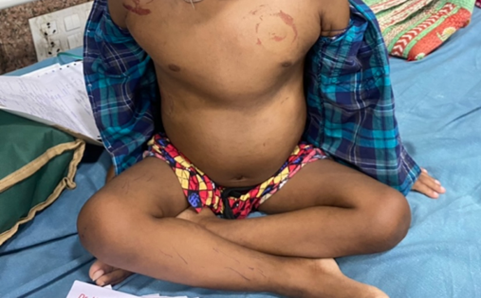
The Case: A child reported with cough and cold for 7 days, with mild fever. He was admitted to the hospital due to breathing difficulties and facial swelling. The clinical exam showed crepitation, wheezing, and pectus carinatum. Patient has history of multiple hospital admissions due to pneumonia and respiratory distress and exposure to tuberculosis. His mother was diagnosed and treated for tuberculosis when he was 3 months old. Condition of the patient was evaluated using ultrasonographic examination, chest radiograph and High-Resolution Computed Tomography of thorax.
Conclusion: High-resolution Computed Tomography (HRCT) scanning is the preferred diagnostic test as it helps to identify the pathologic changes and the exact extent through which it has taken place. Early intervention plays a critical role in reducing severe complications like hemoptysis and cor pulmonale. The current treatment options consist of antibiotics, bronchodilators, anti-inflammatory medications, and physical therapy. The patient was treated using steroids, anti-microbials and inhalational bronchodilators. Complete symptom resolution was noted in two weeks from date of admission. He also seemed to be doing well in the follow-up visit, one week post discharge. Severe cases may require injectable antibiotics. As a widespread condition in India, early diagnosis and treatment with suitable antimicrobials is critical for a positive outcome.
References
Koul PA, Dhar R. 'World Bronchiectasis Day': The Indian perspective. Lung India. 2022;39(4):313-314.
Flume PA, Chalmers JD, Olivier KN. Advances in bronchiectasis: endotyping, genetics, microbiome, and disease heterogeneity. Lancet. 2018 Sep 8;392(10150):880-890.
Kumar V, Abbas AK, Aster JC, Perkins JA. Robbins & Cotran Pathologic Basis of Disease. 10th ed. Philadelphia: Elsevier; 2021.
World Health Organization. Weight- for-age. Available from: https://www.who.int/tools/child-growth-standards/standards/weight-for-age. Last updated Apr 26, 2024; Cited: Oct 26, 2024.
Amati F, Simonetta E, Gramegna A, Tarsia P, Contarini M, Blasi F, et al. The biology of pulmonary exacerbations in bronchiectasis. Eur Respir Rev. 2019;28(154):190055.
Goyal V, Chang AB. Bronchiectasis in Childhood. Clin Chest Med. 2022;43(1):71-88.
Hariprasad K, Krishnan S, Mehta RM. Bronchiectasis in India: Results from the EMBARC and Respiratory Research Network of India Registry. Natl Med J India. 2020;33(2):99-101.
Imam JS, Duarte AG. Non-CF bronchiectasis: Orphan disease no longer. Respir Med. 2020;166:105940.
Wilkinson I. Oxford Handbook of Clinical Medicine. 10th ed. Oxford: Oxford University Press; 2017.
Chapman S. Oxford Handbook of Respiratory Medicine. 4th ed. Oxford: Oxford University Press; 2021

Published
How to Cite
License
Copyright (c) 2024 Anuva Dasgupta, Dibyendu Raychaudhuri

This work is licensed under a Creative Commons Attribution 4.0 International License.
Authors who publish with this journal agree to the following terms:
- The Author retains copyright in the Work, where the term “Work” shall include all digital objects that may result in subsequent electronic publication or distribution.
- Upon acceptance of the Work, the author shall grant to the Publisher the right of first publication of the Work.
- The Author shall grant to the Publisher and its agents the nonexclusive perpetual right and license to publish, archive, and make accessible the Work in whole or in part in all forms of media now or hereafter known under a Creative Commons Attribution 4.0 International License or its equivalent, which, for the avoidance of doubt, allows others to copy, distribute, and transmit the Work under the following conditions:
- Attribution—other users must attribute the Work in the manner specified by the author as indicated on the journal Web site; with the understanding that the above condition can be waived with permission from the Author and that where the Work or any of its elements is in the public domain under applicable law, that status is in no way affected by the license.
- The Author is able to enter into separate, additional contractual arrangements for the nonexclusive distribution of the journal's published version of the Work (e.g., post it to an institutional repository or publish it in a book), as long as there is provided in the document an acknowledgment of its initial publication in this journal.
- Authors are permitted and encouraged to post online a prepublication manuscript (but not the Publisher’s final formatted PDF version of the Work) in institutional repositories or on their Websites prior to and during the submission process, as it can lead to productive exchanges, as well as earlier and greater citation of published work. Any such posting made before acceptance and publication of the Work shall be updated upon publication to include a reference to the Publisher-assigned DOI (Digital Object Identifier) and a link to the online abstract for the final published Work in the Journal.
- Upon Publisher’s request, the Author agrees to furnish promptly to Publisher, at the Author’s own expense, written evidence of the permissions, licenses, and consents for use of third-party material included within the Work, except as determined by Publisher to be covered by the principles of Fair Use.
- The Author represents and warrants that:
- the Work is the Author’s original work;
- the Author has not transferred, and will not transfer, exclusive rights in the Work to any third party;
- the Work is not pending review or under consideration by another publisher;
- the Work has not previously been published;
- the Work contains no misrepresentation or infringement of the Work or property of other authors or third parties; and
- the Work contains no libel, invasion of privacy, or other unlawful matter.
- The Author agrees to indemnify and hold Publisher harmless from the Author’s breach of the representations and warranties contained in Paragraph 6 above, as well as any claim or proceeding relating to Publisher’s use and publication of any content contained in the Work, including third-party content.
Enforcement of copyright
The IJMS takes the protection of copyright very seriously.
If the IJMS discovers that you have used its copyright materials in contravention of the license above, the IJMS may bring legal proceedings against you seeking reparation and an injunction to stop you using those materials. You could also be ordered to pay legal costs.
If you become aware of any use of the IJMS' copyright materials that contravenes or may contravene the license above, please report this by email to contact@ijms.org
Infringing material
If you become aware of any material on the website that you believe infringes your or any other person's copyright, please report this by email to contact@ijms.org


