Case Report: An Atypical Sellar Mass - Sellar Tuberculoma in a Young Patient
DOI:
https://doi.org/10.5195/ijms.2024.2128Keywords:
Intracranial Tuberculoma, pituitary disease, antitubercular agents, Tuberculoma, Sellar Mass, Central Nervous System TB, MRI Imaging, Transsphenoidal Surgery, Pituitary Lesion, Hypopituitarism, Case Report, Anti-Tubercular Therapy, Endocrine DysfunctionAbstract
Background: Tuberculosis of the central nervous system is an uncommon but one of the most severe forms. It manifests as tuberculoma and tuberculous meningitis, with the majority of cases affecting children and immunocompromised patients. Overall, tuberculomas make up to 0.15–2 % of all intracranial lesions but sellar tuberculoma is extremely rare.
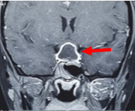
The Case: An 18-year-old female patient presented with complaint of generalized weakness, eye pain and headache for 3-4 months. Magnetic resonance imaging (MRI) of brain showed sellar and suprasellar space occupying lesion. Trans sphenoidal approach was used to remove the lesion completely. A sellar tuberculoma was confirmed on pathological evaluation and the patient was put on postoperative anti-tubercular therapy.
Conclusion: Although rare, intracranial tuberculomas, particularly those that originate in the sellar, are notorious for mimicking pituitary tumors by jeopardizing pituitary hormonal function and applying compressive forces on surrounding intracranial structures. However, a prompt assessment can help overcome this diagnostic difficulty with the timely initiation of anti-tubercular therapy (ATT).
References
Global Tuberculosis Report 2022. Geneva: World Health Organization; 2022.
DeLance AR, Safaee M, Oh MC, Clark AJ, Kaur G, Sun MZ, et al. Tuberculoma of the central nervous system. J Clin Neurosci. 2013;20(10):1333-41.
Ayele B, Wako A, Shewaye A, Tessema A. Sellar Tuberculoma: A rare presentation in a 30-year-old Ethiopian woman: case report. Afr J Neurol Sci. 2019;38(1):50-53.
Dastur HM. Tuberculoma. In: Vinken PJ, Bruyn GW, editors. Handbook of Clinical Neurology, vol. 18. New York: American Elsevier; 1975:413–26.
DeAngelis LM. Intracranial tuberculoma: case report and review of the literature. Neurology. 1981;31(9):1133-6.
Maurice-Williams RS. Tuberculomas of the brain in Britain. Postgrad Med J. 1972;48(565):679-81.
Ranjan A, Chandy MJ. Intrasellar tuberculoma. Br J Neurosurg. 1994;8(2):179-85.
Kumar T, Nigam JS, Jamal I, Jha VC. Primary pituitary tuberculosis. Autops Case Rep. 2020;11: e2020228.
Furtado SV, Venkatesh PK, Ghosal N, Hegde AS. Isolated sellar tuberculoma presenting with panhypopituitarism: clinical, diagnostic considerations and literature review. Neurol Sci. 2011;32(2):301-4.
Pereira J, Vaz R, Carvalho D, Cruz C. Thickening of the pituitary stalk: a finding suggestive of intrasellar tuberculoma? Case report. Neurosurgery. 1995;36(5):1013-5; discussion 1015-6.
Prabha BB, Rangachari V, Subramaniam V, Gopan TV, Baliga VB. Pituitary tuberculoma masquerading as a pituitary adenoma: interesting case report and review of literature. Asian J Neurosurg. 2021;16(1):141-3.
Dutta P, Bhansali A, Singh P, Bhat MH. Suprasellar tubercular abscess presenting as panhypopituitarism: a common lesion in an uncommon site with a brief review of literature. Pituitary 2006;9(1):73-7
Ben Abid F, Abukhattab M, Karim H, Agab M, Al-Bozom I, Ibrahim WH. Primary pituitary tuberculosis revisited. Am J Case Rep. 2017;18:391-4.

Published
How to Cite
License
Copyright (c) 2024 Arwa Jamali, Rakeshkumar Luhana

This work is licensed under a Creative Commons Attribution 4.0 International License.
Authors who publish with this journal agree to the following terms:
- The Author retains copyright in the Work, where the term “Work” shall include all digital objects that may result in subsequent electronic publication or distribution.
- Upon acceptance of the Work, the author shall grant to the Publisher the right of first publication of the Work.
- The Author shall grant to the Publisher and its agents the nonexclusive perpetual right and license to publish, archive, and make accessible the Work in whole or in part in all forms of media now or hereafter known under a Creative Commons Attribution 4.0 International License or its equivalent, which, for the avoidance of doubt, allows others to copy, distribute, and transmit the Work under the following conditions:
- Attribution—other users must attribute the Work in the manner specified by the author as indicated on the journal Web site; with the understanding that the above condition can be waived with permission from the Author and that where the Work or any of its elements is in the public domain under applicable law, that status is in no way affected by the license.
- The Author is able to enter into separate, additional contractual arrangements for the nonexclusive distribution of the journal's published version of the Work (e.g., post it to an institutional repository or publish it in a book), as long as there is provided in the document an acknowledgment of its initial publication in this journal.
- Authors are permitted and encouraged to post online a prepublication manuscript (but not the Publisher’s final formatted PDF version of the Work) in institutional repositories or on their Websites prior to and during the submission process, as it can lead to productive exchanges, as well as earlier and greater citation of published work. Any such posting made before acceptance and publication of the Work shall be updated upon publication to include a reference to the Publisher-assigned DOI (Digital Object Identifier) and a link to the online abstract for the final published Work in the Journal.
- Upon Publisher’s request, the Author agrees to furnish promptly to Publisher, at the Author’s own expense, written evidence of the permissions, licenses, and consents for use of third-party material included within the Work, except as determined by Publisher to be covered by the principles of Fair Use.
- The Author represents and warrants that:
- the Work is the Author’s original work;
- the Author has not transferred, and will not transfer, exclusive rights in the Work to any third party;
- the Work is not pending review or under consideration by another publisher;
- the Work has not previously been published;
- the Work contains no misrepresentation or infringement of the Work or property of other authors or third parties; and
- the Work contains no libel, invasion of privacy, or other unlawful matter.
- The Author agrees to indemnify and hold Publisher harmless from the Author’s breach of the representations and warranties contained in Paragraph 6 above, as well as any claim or proceeding relating to Publisher’s use and publication of any content contained in the Work, including third-party content.
Enforcement of copyright
The IJMS takes the protection of copyright very seriously.
If the IJMS discovers that you have used its copyright materials in contravention of the license above, the IJMS may bring legal proceedings against you seeking reparation and an injunction to stop you using those materials. You could also be ordered to pay legal costs.
If you become aware of any use of the IJMS' copyright materials that contravenes or may contravene the license above, please report this by email to contact@ijms.org
Infringing material
If you become aware of any material on the website that you believe infringes your or any other person's copyright, please report this by email to contact@ijms.org


