
A Case of Successful Surgical Resection of Locally Advanced (T4) Lung Cancer Utilizing a Multi-Disciplinary Approach Involving Previously Unresectable Structures
DOI:
https://doi.org/10.5195/ijms.2025.2516Keywords:
lung cancer, cardiothoracic surgery, T4, locally advanced, surgeryAbstract
Background:
Lung cancer is the leading cause of cancer death worldwide and the second most prevalent cancer in the world.(1) Tumor node metastasis (TNM) staging continues to serve as the primary prognostic factor for survival in lung cancer.
TNM classification (8th edition) characterizes T4 disease as a tumor exceeding 7 cm in its largest dimension or one that invades structures such as the mediastinum, diaphragm, heart, great vessels, recurrent laryngeal nerve, carina, trachea, esophagus, spine or represents a separate tumor in a different lobe of ipsilateral lung.(2) These structures are typically deemed “unresectable”.(3)
Case:
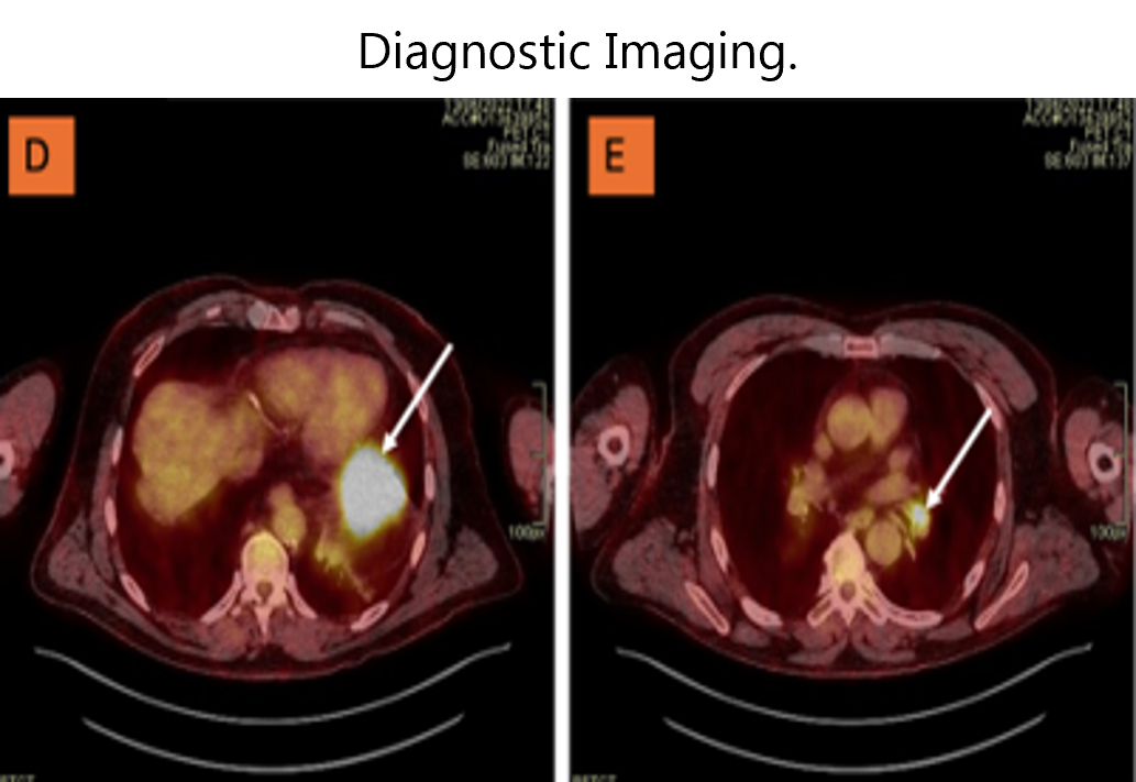
We present the case of an asymptomatic 66-year-old gentleman with an incidental lung nodule found on routine pre-operative chest x ray. In this case, imaging revealed local extension, involving the stomach (excluded from TNM classification), diaphragm and pericardium. Consequently, the disease was initially considered incurable. Nevertheless, through collaborative multi-disciplinary surgical approach, successful resection was achieved.
Conclusion:
T4 disease exhibits heterogeneity, and although it is typically deemed unresectable, recent developments in surgery are challenging this conventional belief and demonstrating the potential benefits of surgical resection, particularly where a radical dissection is anticipated.
References
Thandra KC, Barsouk A, Saginala K, Aluru JS, Barsouk A. Epidemiology of lung cancer. Contemp Oncol (Pozn). 2021;25(1):45–52.
Lababede O, Meziane MA. The eighth edition of TNM staging of lung cancer: Reference chart and diagrams. Oncologist. 2018;23(7):844–848.
Erasmus LT, Strange CD, Ahuja J, Agrawal R, Shroff GS, Marom EM, et al. Imaging of lung cancer staging: TNM 9 updates. Semin Ultrasound CT MR. 2024.
Dartevelle P, Mitilian D, Fadel E. Extended surgery for T4 lung cancer: A 30 years’ experience. Gen Thorac Cardiovasc Surg. 2017;65:321–328.
Aldarouish M, Wang C. Trends and advances in tumor immunology and lung cancer immunotherapy. J Exp Clin Cancer Res. 2016;35(1):1–13.
DiPerna CA, Wood DE. Surgical management of T3 and T4 lung cancer. Clin Cancer Res. 2005;11(13):5038s–5044s.
Port JL, Korst RJ, Lee PC, Kansler AL, Kerem Y, Altorki NK. Surgical resection for multifocal (T4) non-small cell lung cancer: Is the T4 designation valid? Ann Thorac Surg. 2007;83(2):397–400.
Rami-Porta R, Bolejack V, Crowley J, Ball D, Kim J, Lyons G, et al. The IASLC lung cancer staging project: Proposals for the revisions of the T descriptors in the forthcoming eighth edition of the TNM classification for lung cancer. J Thorac Oncol. 2015;10(7):990–1003.
Jung HS, Lee JG, Lee CY, Kim DJ, Chung KY. Validation of the T descriptor in the new 8th TNM classification for non-small cell lung cancer. J Thorac Dis. 2018;10(1):162.
Tankel J, Mouhanna J, Katz A, Fiset P-O, Rayes R, Siblini A, et al. The 8th edition TNM stage reclassification of T4 non-small cell lung cancer: A granular examination of short and long-term outcomes. Clin Lung Cancer. 2023.
Yamanashi K, Menju T, Hamaji M, Tanaka S, Yutaka Y, Yamada Y, et al. Prognostic factors related to postoperative survival in the newly classified clinical T4 lung cancer. Eur J Cardiothorac Surg. 2020;57(4):754–761.
Grunenwald DH, André F, Le Péchoux C, Girard P, Lamer C, Laplanche A, et al. Benefit of surgery after chemoradiotherapy in stage IIIB (T4 and/or N3) non–small cell lung cancer. J Thorac Cardiovasc Surg. 2001;122(4):796–802.
Verdecchia A, Francisci S, Brenner H, Gatta G, Micheli A, Mangone L, et al. Recent cancer survival in Europe: A 2000–02 period analysis of EUROCARE-4 data. Lancet Oncol. 2007;8(9):784–796.
Brown JK, Singh K, Dumitru R, Chan E, Kim MP. The benefits of enhanced recovery after surgery programs and their application in cardiothoracic surgery. Methodist Debakey Cardiovasc J. 2018;14(2):77.
Macchiarini P, Chapelier AR, Monnet I, Vannetzel J-M, Rebischung J-L, Cerrina J, et al. Extended operations after induction therapy for stage IIIb (T4) non-small cell lung cancer. Ann Thorac Surg. 1994;57(4):966–973.
Furrer K, Weder W, Eboulet EI, Betticher D, Pless M, Stupp R, et al. Extended resection for potentially operable patients with stage III non-small cell lung cancer after induction treatment. J Thorac Cardiovasc Surg. 2022;164(6):1587–1602.e5.
Pignon J-P, Tribodet H, Scagliotti GV, Douillard J-Y, Shepherd FA, Stephens RJ, et al. Lung adjuvant cisplatin evaluation: A pooled analysis by the LACE Collaborative Group. J Clin Oncol. 2008;26(21):3552–3559
Published
How to Cite
Issue
Section
License
Copyright (c) 2025 Meadhbh Ni Mhiochain de Grae, William P. Duggan Duggan , Paul A. Carroll , Alan Soo

This work is licensed under a Creative Commons Attribution 4.0 International License.
Authors who publish with this journal agree to the following terms:
- The Author retains copyright in the Work, where the term “Work” shall include all digital objects that may result in subsequent electronic publication or distribution.
- Upon acceptance of the Work, the author shall grant to the Publisher the right of first publication of the Work.
- The Author shall grant to the Publisher and its agents the nonexclusive perpetual right and license to publish, archive, and make accessible the Work in whole or in part in all forms of media now or hereafter known under a Creative Commons Attribution 4.0 International License or its equivalent, which, for the avoidance of doubt, allows others to copy, distribute, and transmit the Work under the following conditions:
- Attribution—other users must attribute the Work in the manner specified by the author as indicated on the journal Web site; with the understanding that the above condition can be waived with permission from the Author and that where the Work or any of its elements is in the public domain under applicable law, that status is in no way affected by the license.
- The Author is able to enter into separate, additional contractual arrangements for the nonexclusive distribution of the journal's published version of the Work (e.g., post it to an institutional repository or publish it in a book), as long as there is provided in the document an acknowledgment of its initial publication in this journal.
- Authors are permitted and encouraged to post online a prepublication manuscript (but not the Publisher’s final formatted PDF version of the Work) in institutional repositories or on their Websites prior to and during the submission process, as it can lead to productive exchanges, as well as earlier and greater citation of published work. Any such posting made before acceptance and publication of the Work shall be updated upon publication to include a reference to the Publisher-assigned DOI (Digital Object Identifier) and a link to the online abstract for the final published Work in the Journal.
- Upon Publisher’s request, the Author agrees to furnish promptly to Publisher, at the Author’s own expense, written evidence of the permissions, licenses, and consents for use of third-party material included within the Work, except as determined by Publisher to be covered by the principles of Fair Use.
- The Author represents and warrants that:
- the Work is the Author’s original work;
- the Author has not transferred, and will not transfer, exclusive rights in the Work to any third party;
- the Work is not pending review or under consideration by another publisher;
- the Work has not previously been published;
- the Work contains no misrepresentation or infringement of the Work or property of other authors or third parties; and
- the Work contains no libel, invasion of privacy, or other unlawful matter.
- The Author agrees to indemnify and hold Publisher harmless from the Author’s breach of the representations and warranties contained in Paragraph 6 above, as well as any claim or proceeding relating to Publisher’s use and publication of any content contained in the Work, including third-party content.
Enforcement of copyright
The IJMS takes the protection of copyright very seriously.
If the IJMS discovers that you have used its copyright materials in contravention of the license above, the IJMS may bring legal proceedings against you seeking reparation and an injunction to stop you using those materials. You could also be ordered to pay legal costs.
If you become aware of any use of the IJMS' copyright materials that contravenes or may contravene the license above, please report this by email to contact@ijms.org
Infringing material
If you become aware of any material on the website that you believe infringes your or any other person's copyright, please report this by email to contact@ijms.org


